2
views
0
recommends
+1 Recommend
2 collections
    0
    shares
      • Record: found
      • Abstract: found
      • Article: found
      Is Open Access

      Spectral-domain optical coherence tomography assessment of retinal and choroidal changes in patients with coronavirus disease 2019: a case-control study

      research-article

      Read this article at

      Bookmark
          There is no author summary for this article yet. Authors can add summaries to their articles on ScienceOpen to make them more accessible to a non-specialist audience.

          Abstract

          Objectives

          This study aimed to evaluate the retinal and choroidal changes in the macular region of patients with Coronavirus Disease 2019 (COVID-19) using structural spectral-domain optical coherence tomography (SD-OCT) analysis.

          Methods

          This cross-sectional observational case-control study included patients recovered from COVID-19. The COVID-19 in all participants was confirmed using the reverse transcription-polymerase chain reaction (RT-PCR) technique. The participants had mild to moderate degree of disease without a history of hospitalization, steroid usage, or blood saturation below 92%. Macular SD-OCT was performed at least two weeks and up to one month after recovery from systemic COVID-19. Quantitative and qualitative changes detected by macular SD-OCT imaging were evaluated in COVID-19 recovered patients and compared with the results of age-matched normal controls.

          Results

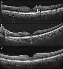
          Participants in this study included 30 cases (60 eyes) and 60 healthy controls (120 eyes). In total, 17 (28.3%) eyes in patient group showed at least one abnormal finding indicated by macular SD-OCT imaging included hyperreflective lesions in different retinal layers. In addition, dilated choroidal vessels and retinal pigment epitheliopathy were evident in 41 (68.3.6%) and 4 (6.6%) eyes in patient group, respectively, and their OCT findings resembled those with pachychoroid spectrum. No statistically significant differences were observed in retinal layers or retinal volume between the two groups. The mean ± SD subfoveal choroidal thickness (SFCT) was determined at 380.3 ± 12.40 μm, which was significantly thicker than that in control group (310.7 ± 57.5 μm) ( P < 0.001).

          Conclusion

          Regarding retinal thickness, no significant change was observed in different retina layers of patients with COVID-19; however, there were striking qualitative changes, such as hyperreflective lesions in different retinal layers. The evaluation of choroidal structure and thickness demonstrated remarkable abnormal pachyvessels and significant thickening of the SFCT but the clinical significance of these findings is unknown.

          Related collections

          Most cited references36

          • Record: found
          • Abstract: found
          • Article: not found

          Tissue distribution of ACE2 protein, the functional receptor for SARS coronavirus. A first step in understanding SARS pathogenesis

          Abstract Severe acute respiratory syndrome (SARS) is an acute infectious disease that spreads mainly via the respiratory route. A distinct coronavirus (SARS‐CoV) has been identified as the aetiological agent of SARS. Recently, a metallopeptidase named angiotensin‐converting enzyme 2 (ACE2) has been identified as the functional receptor for SARS‐CoV. Although ACE2 mRNA is known to be present in virtually all organs, its protein expression is largely unknown. Since identifying the possible route of infection has major implications for understanding the pathogenesis and future treatment strategies for SARS, the present study investigated the localization of ACE2 protein in various human organs (oral and nasal mucosa, nasopharynx, lung, stomach, small intestine, colon, skin, lymph nodes, thymus, bone marrow, spleen, liver, kidney, and brain). The most remarkable finding was the surface expression of ACE2 protein on lung alveolar epithelial cells and enterocytes of the small intestine. Furthermore, ACE2 was present in arterial and venous endothelial cells and arterial smooth muscle cells in all organs studied. In conclusion, ACE2 is abundantly present in humans in the epithelia of the lung and small intestine, which might provide possible routes of entry for the SARS‐CoV. This epithelial expression, together with the presence of ACE2 in vascular endothelium, also provides a first step in understanding the pathogenesis of the main SARS disease manifestations. Copyright © 2004 Pathological Society of Great Britain and Ireland. Published by John Wiley & Sons, Ltd.
            Bookmark
            • Record: found
            • Abstract: found
            • Article: not found

            Receptor Recognition by the Novel Coronavirus from Wuhan: an Analysis Based on Decade-Long Structural Studies of SARS Coronavirus

            The recent emergence of Wuhan coronavirus (2019-nCoV) puts the world on alert. 2019-nCoV is reminiscent of the SARS-CoV outbreak in 2002 to 2003. Our decade-long structural studies on the receptor recognition by SARS-CoV have identified key interactions between SARS-CoV spike protein and its host receptor angiotensin-converting enzyme 2 (ACE2), which regulate both the cross-species and human-to-human transmissions of SARS-CoV. One of the goals of SARS-CoV research was to build an atomic-level iterative framework of virus-receptor interactions to facilitate epidemic surveillance, predict species-specific receptor usage, and identify potential animal hosts and animal models of viruses. Based on the sequence of 2019-nCoV spike protein, we apply this predictive framework to provide novel insights into the receptor usage and likely host range of 2019-nCoV. This study provides a robust test of this reiterative framework, providing the basic, translational, and public health research communities with predictive insights that may help study and battle this novel 2019-nCoV.
              Bookmark
              • Record: found
              • Abstract: found
              • Article: not found

              Can the Coronavirus Disease 2019 (COVID-19) Affect the Eyes? A Review of Coronaviruses and Ocular Implications in Humans and Animals

              ABSTRACT In December 2019, a novel coronavirus (CoV) epidemic, caused by the severe acute respiratory syndrome coronavirus – 2 (SARS-CoV-2) emerged from China. This virus causes the coronavirus disease 2019 (COVID-19). Since then, there have been anecdotal reports of ocular infection. The ocular implications of human CoV infections have not been widely studied. However, CoVs have been known to cause various ocular infections in animals. Clinical entities such as conjunctivitis, anterior uveitis, retinitis, and optic neuritis have been documented in feline and murine models. In this article, the current evidence suggesting possible human CoV infection of ocular tissue is reviewed. The review article will also highlight animal CoVs and their associated ocular infections. We hope that this article will serve as a start for further research into the ocular implications of human CoV infections.
                Bookmark

                Author and article information

                Contributors
                m_ansariastaneh@yahoo.com
                Journal
                J Ophthalmic Inflamm Infect
                J Ophthalmic Inflamm Infect
                Journal of Ophthalmic Inflammation and Infection
                Springer Berlin Heidelberg (Berlin/Heidelberg )
                1869-5760
                18 June 2022
                18 June 2022
                2022
                : 12
                : 18
                Affiliations
                [1 ]GRID grid.411583.a, ISNI 0000 0001 2198 6209, Eye Research Center, , Mashhad University of Medical Sciences, ; Mashhad, Iran
                [2 ]GRID grid.411583.a, ISNI 0000 0001 2198 6209, Department of Internal Medicine, Faculty of Medicine, , Mashhad University of Medical Sciences, ; Mashhad, Iran
                [3 ]GRID grid.411583.a, ISNI 0000 0001 2198 6209, Department of Medical Informatics, Faculty of Medicine, , Mashhad University of Medical Sciences, ; Mashhad, Iran
                [4 ]GRID grid.411583.a, ISNI 0000 0001 2198 6209, Pharmaceutical Research Center, School of Pharmacy, , Mashhad University of Medical Sciences, ; Mashhad, Iran
                Article
                297
                10.1186/s12348-022-00297-z
                9206085
                35716213
                d25bcd89-06dc-4df2-9a4f-0f539d65438a
                © The Author(s) 2022

                Open AccessThis article is licensed under a Creative Commons Attribution 4.0 International License, which permits use, sharing, adaptation, distribution and reproduction in any medium or format, as long as you give appropriate credit to the original author(s) and the source, provide a link to the Creative Commons licence, and indicate if changes were made. The images or other third party material in this article are included in the article's Creative Commons licence, unless indicated otherwise in a credit line to the material. If material is not included in the article's Creative Commons licence and your intended use is not permitted by statutory regulation or exceeds the permitted use, you will need to obtain permission directly from the copyright holder. To view a copy of this licence, visit http://creativecommons.org/licenses/by/4.0/.

                History
                : 5 November 2021
                : 2 June 2022
                Funding
                Funded by: Vice-Chancellor of Research, Mashhad University of Medical Sciences, Mashhad
                Award ID: 990069
                Award Recipient :
                Categories
                Original Research
                Custom metadata
                © The Author(s) 2022

                Ophthalmology & Optometry
                choroid,coronavirus disease 2019,macula,retina,severe acute respiratory syndrome corona virus 2,spectral-domain optical coherence tomography

                Comments

                Comment on this article