45
views
0
recommends
+1 Recommend
1 collections
    0
    shares
      • Record: found
      • Abstract: found
      • Article: found
      Is Open Access

      Senolytics reduce coronavirus-related mortality in old mice

      research-article
      1 , 1 , 2 , 3 , 2 , 4 , 4 , 1 , 1 , 2 , 5 , 5 , 2 , 2 , 2 , 2 , 2 , 6 , 2 , 2 , 1 , 7 , 8 , 2 , 9 , 2 , 10 , 7 , 8 , 5 , 4 , 11 , 11 , 3 , 12 , 13 , 4 , * , 2 , 3 , * , 1 , * , 2 , 3 , 14 , * , 1 , *
      Science (New York, N.Y.)

      Read this article at

      Bookmark
          There is no author summary for this article yet. Authors can add summaries to their articles on ScienceOpen to make them more accessible to a non-specialist audience.

          Abstract

          INTRODUCTION:

          The COVID-19 pandemic revealed enhanced vulnerability of the elderly and chronically ill to adverse outcomes upon severe acute respiratory syndrome coronavirus 2 (SARS-CoV-2) infection. Senescence is a cell fate elicited by cellular stress that results in changes in gene expression, morphology, metabolism, and resistance to apoptosis. Senescent cells (SnCs) secrete pro-inflammatory factors, called the senescence-associated secretory phenotype (SASP). SnCs accumulate with age and drive chronic inflammation. In human cells and tissues and using a new infection paradigm, we asked whether SnCs are a cause of adverse outcomes of infection with aging. This is relevant because SnCs can be selectively eliminated in vivo with a new class of therapeutics called senolytics, potentially affording a new approach to treat COVID-19.

          RATIONALE:

          We hypothesized that SnCs, because of their pro-inflammatory SASP, might have a heightened response to pathogen-associated molecular pattern (PAMP) factors, resulting in increased risk of cytokine storm and multi-organ failure. To test this, we treated senescent and nonsenescent human cells with the PAMPs lipopolysaccharide (LPS) and SARS-CoV-2 spike protein (S1) and measured the SASP and its effect on non-SnCs. Similarly, old and progeroid mice were challenged with LPS, and we measured the SASP. Previously, we created a “normal microbial experience” (NME) for mice by transmitting environmental pathogens to specified-pathogen–free (SPF) mice through exposure to pet store mice or their bedding. The first pathogen transferred was mouse hepatitis virus (MHV), a β-coronavirus closely related to SARS-CoV-2. NME rapidly killed aged SPF mice known to have an increased burden of SnCs compared with young SPF mice, which survive NME. This afforded an experimental paradigm to test whether senolytics blunt adverse outcomes in β-coronavirus infection.

          RESULTS:

          Human endothelial SnCs became hyperinflammatory in response to challenge with LPS and S1, relative to non-SnCs. The PAMP-elicited secretome of SnCs caused increased expression of viral entry proteins and reduced expression of antiviral genes in non-senescent human endothelial and lung epithelial cells, and the proximity of these events was established in human lung biopsies. Treatment of old mice with LPS significantly increased SASP expression in several organs relative to young mice, confirming our hypothesis in vivo. Similarly, old mice exposed to NME displayed a significant multi-organ increase in SnCs and the SASP, impaired immune response to MHV, and 100% mortality, whereas inoculation with antibodies against MHV before NME afforded complete rescue of mortality. Treating old mice with the senolytic fisetin, which selectively eliminates SnCs after NME reduced mortality by 50%, reduced expression of inflammatory proteins in serum and tissue and improved the immune response. This was confirmed with a second senolytic regimen, Dasatinib plus Quercetin, as well as genetic ablation of SnCs in aged mice, establishing SnCs as a cause of adverse outcomes in aged organisms exposed to a new viral pathogen.

          CONCLUSION:

          SnCs amplify susceptibility to COVID-19 and pathogen-induced hyperinflammation. Reducing SnC burden in aged mice reduces mortality after pathogen exposure, including a β-coronavirus. Our findings strongly support the Geroscience hypothesis that therapeutically targeting fundamental aging mechanisms improves resilience in the elderly, with alleviation of morbidity and mortality due to pathogenic stress. This suggests that senolytics might protect others vulnerable to adverse COVID-19 outcomes in whom increased SnCs occur (such as in obesity or numerous chronic diseases).

          Graphical Abstract

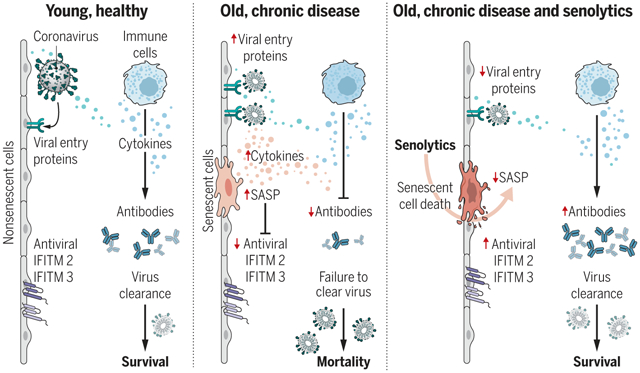
          SnCs that accumulate with age or chronic disease react to PAMPs such as SARS-CoV-2 S1 by amplifying the SASP, which increases viral entry protein expression and decreases viral defense IFITMs in normal cells. Old mice exposed to pathogens such as the β-coronavirus MHV have increased inflammation and higher mortality. Treatment with a senolytic decreased SnCs, inflammation, and mortality and increased the antiviral antibody response.

          Abstract

          The COVID-19 pandemic has revealed the pronounced vulnerability of the elderly and chronically ill to severe acute respiratory syndrome coronavirus 2 (SARS-CoV-2)–induced morbidity and mortality. Cellular senescence contributes to inflammation, multiple chronic diseases, and age-related dysfunction, but effects on responses to viral infection are unclear. Here, we demonstrate that senescent cells (SnCs) become hyper-inflammatory in response to pathogen-associated molecular patterns (PAMPs), including SARS-CoV-2 spike protein-1, increasing expression of viral entry proteins and reducing antiviral gene expression in non-SnCs through a paracrine mechanism. Old mice acutely infected with pathogens that included a SARS-CoV-2–related mouse β-coronavirus experienced increased senescence and inflammation, with nearly 100% mortality. Targeting SnCs by using senolytic drugs before or after pathogen exposure significantly reduced mortality, cellular senescence, and inflammatory markers and increased antiviral antibodies. Thus, reducing the SnC burden in diseased or aged individuals should enhance resilience and reduce mortality after viral infection, including that of SARS-CoV-2.

          Related collections

          Most cited references59

          • Record: found
          • Abstract: found
          • Article: not found

          Clinical features of patients infected with 2019 novel coronavirus in Wuhan, China

          Summary Background A recent cluster of pneumonia cases in Wuhan, China, was caused by a novel betacoronavirus, the 2019 novel coronavirus (2019-nCoV). We report the epidemiological, clinical, laboratory, and radiological characteristics and treatment and clinical outcomes of these patients. Methods All patients with suspected 2019-nCoV were admitted to a designated hospital in Wuhan. We prospectively collected and analysed data on patients with laboratory-confirmed 2019-nCoV infection by real-time RT-PCR and next-generation sequencing. Data were obtained with standardised data collection forms shared by WHO and the International Severe Acute Respiratory and Emerging Infection Consortium from electronic medical records. Researchers also directly communicated with patients or their families to ascertain epidemiological and symptom data. Outcomes were also compared between patients who had been admitted to the intensive care unit (ICU) and those who had not. Findings By Jan 2, 2020, 41 admitted hospital patients had been identified as having laboratory-confirmed 2019-nCoV infection. Most of the infected patients were men (30 [73%] of 41); less than half had underlying diseases (13 [32%]), including diabetes (eight [20%]), hypertension (six [15%]), and cardiovascular disease (six [15%]). Median age was 49·0 years (IQR 41·0–58·0). 27 (66%) of 41 patients had been exposed to Huanan seafood market. One family cluster was found. Common symptoms at onset of illness were fever (40 [98%] of 41 patients), cough (31 [76%]), and myalgia or fatigue (18 [44%]); less common symptoms were sputum production (11 [28%] of 39), headache (three [8%] of 38), haemoptysis (two [5%] of 39), and diarrhoea (one [3%] of 38). Dyspnoea developed in 22 (55%) of 40 patients (median time from illness onset to dyspnoea 8·0 days [IQR 5·0–13·0]). 26 (63%) of 41 patients had lymphopenia. All 41 patients had pneumonia with abnormal findings on chest CT. Complications included acute respiratory distress syndrome (12 [29%]), RNAaemia (six [15%]), acute cardiac injury (five [12%]) and secondary infection (four [10%]). 13 (32%) patients were admitted to an ICU and six (15%) died. Compared with non-ICU patients, ICU patients had higher plasma levels of IL2, IL7, IL10, GSCF, IP10, MCP1, MIP1A, and TNFα. Interpretation The 2019-nCoV infection caused clusters of severe respiratory illness similar to severe acute respiratory syndrome coronavirus and was associated with ICU admission and high mortality. Major gaps in our knowledge of the origin, epidemiology, duration of human transmission, and clinical spectrum of disease need fulfilment by future studies. Funding Ministry of Science and Technology, Chinese Academy of Medical Sciences, National Natural Science Foundation of China, and Beijing Municipal Science and Technology Commission.
            Bookmark
            • Record: found
            • Abstract: found
            • Article: found

            COVID-19: consider cytokine storm syndromes and immunosuppression

            As of March 12, 2020, coronavirus disease 2019 (COVID-19) has been confirmed in 125 048 people worldwide, carrying a mortality of approximately 3·7%, 1 compared with a mortality rate of less than 1% from influenza. There is an urgent need for effective treatment. Current focus has been on the development of novel therapeutics, including antivirals and vaccines. Accumulating evidence suggests that a subgroup of patients with severe COVID-19 might have a cytokine storm syndrome. We recommend identification and treatment of hyperinflammation using existing, approved therapies with proven safety profiles to address the immediate need to reduce the rising mortality. Current management of COVID-19 is supportive, and respiratory failure from acute respiratory distress syndrome (ARDS) is the leading cause of mortality. 2 Secondary haemophagocytic lymphohistiocytosis (sHLH) is an under-recognised, hyperinflammatory syndrome characterised by a fulminant and fatal hypercytokinaemia with multiorgan failure. In adults, sHLH is most commonly triggered by viral infections 3 and occurs in 3·7–4·3% of sepsis cases. 4 Cardinal features of sHLH include unremitting fever, cytopenias, and hyperferritinaemia; pulmonary involvement (including ARDS) occurs in approximately 50% of patients. 5 A cytokine profile resembling sHLH is associated with COVID-19 disease severity, characterised by increased interleukin (IL)-2, IL-7, granulocyte-colony stimulating factor, interferon-γ inducible protein 10, monocyte chemoattractant protein 1, macrophage inflammatory protein 1-α, and tumour necrosis factor-α. 6 Predictors of fatality from a recent retrospective, multicentre study of 150 confirmed COVID-19 cases in Wuhan, China, included elevated ferritin (mean 1297·6 ng/ml in non-survivors vs 614·0 ng/ml in survivors; p 39·4°C 49 Organomegaly None 0 Hepatomegaly or splenomegaly 23 Hepatomegaly and splenomegaly 38 Number of cytopenias * One lineage 0 Two lineages 24 Three lineages 34 Triglycerides (mmol/L) 4·0 mmol/L 64 Fibrinogen (g/L) >2·5 g/L 0 ≤2·5 g/L 30 Ferritin ng/ml 6000 ng/ml 50 Serum aspartate aminotransferase <30 IU/L 0 ≥30 IU/L 19 Haemophagocytosis on bone marrow aspirate No 0 Yes 35 Known immunosuppression † No 0 Yes 18 The Hscore 11 generates a probability for the presence of secondary HLH. HScores greater than 169 are 93% sensitive and 86% specific for HLH. Note that bone marrow haemophagocytosis is not mandatory for a diagnosis of HLH. HScores can be calculated using an online HScore calculator. 11 HLH=haemophagocytic lymphohistiocytosis. * Defined as either haemoglobin concentration of 9·2 g/dL or less (≤5·71 mmol/L), a white blood cell count of 5000 white blood cells per mm3 or less, or platelet count of 110 000 platelets per mm3 or less, or all of these criteria combined. † HIV positive or receiving longterm immunosuppressive therapy (ie, glucocorticoids, cyclosporine, azathioprine).
              Bookmark
              • Record: found
              • Abstract: found
              • Article: not found

              Clinical predictors of mortality due to COVID-19 based on an analysis of data of 150 patients from Wuhan, China

              Dear Editor, The rapid emergence of COVID-19 in Wuhan city, Hubei Province, China, has resulted in thousands of deaths [1]. Many infected patients, however, presented mild flu-like symptoms and quickly recover [2]. To effectively prioritize resources for patients with the highest risk, we identified clinical predictors of mild and severe patient outcomes. Using the database of Jin Yin-tan Hospital and Tongji Hospital, we conducted a retrospective multicenter study of 68 death cases (68/150, 45%) and 82 discharged cases (82/150, 55%) with laboratory-confirmed infection of SARS-CoV-2. Patients met the discharge criteria if they had no fever for at least 3 days, significantly improved respiratory function, and had negative SARS-CoV-2 laboratory test results twice in succession. Case data included demographics, clinical characteristics, laboratory results, treatment options and outcomes. For statistical analysis, we represented continuous measurements as means (SDs) or as medians (IQRs) which compared with Student’s t test or the Mann–Whitney–Wilcoxon test. Categorical variables were expressed as numbers (%) and compared by the χ 2 test or Fisher’s exact test. The distribution of the enrolled patients’ age is shown in Fig. 1a. There was a significant difference in age between the death group and the discharge group (p < 0.001) but no difference in the sex ratio (p = 0.43). A total of 63% (43/68) of patients in the death group and 41% (34/82) in the discharge group had underlying diseases (p = 0.0069). It should be noted that patients with cardiovascular diseases have a significantly increased risk of death when they are infected with SARS-CoV-2 (p < 0.001). A total of 16% (11/68) of the patients in the death group had secondary infections, and 1% (1/82) of the patients in the discharge group had secondary infections (p = 0.0018). Laboratory results showed that there were significant differences in white blood cell counts, absolute values of lymphocytes, platelets, albumin, total bilirubin, blood urea nitrogen, blood creatinine, myoglobin, cardiac troponin, C-reactive protein (CRP) and interleukin-6 (IL-6) between the two groups (Fig. 1b and Supplementary Table 1). Fig. 1 a Age distribution of patients with confirmed COVID-19; b key laboratory parameters for the outcomes of patients with confirmed COVID-19; c interval from onset of symptom to death of patients with confirmed COVID-19; d summary of the cause of death of 68 died patients with confirmed COVID-19 The survival times of the enrolled patients in the death group were analyzed. The distribution of survival time from disease onset to death showed two peaks, with the first one at approximately 14 days (22 cases) and the second one at approximately 22 days (17 cases) (Fig. 1c). An analysis of the cause of death was performed. Among the 68 fatal cases, 36 patients (53%) died of respiratory failure, five patients (7%) with myocardial damage died of circulatory failure, 22 patients (33%) died of both, and five remaining died of an unknown cause (Fig. 1d). Based on the analysis of the clinical data, we confirmed that some patients died of fulminant myocarditis. In this study, we first reported that the infection of SARS-CoV-2 may cause fulminant myocarditis. Given that fulminant myocarditis is characterized by a rapid progress and a severe state of illness [3], our results should alert physicians to pay attention not only to the symptoms of respiratory dysfunction but also the symptoms of cardiac injury. Further, large-scale studies and the studies on autopsy are needed to confirm our analysis. In conclusion, predictors of a fatal outcome in COVID-19 cases included age, the presence of underlying diseases, the presence of secondary infection and elevated inflammatory indicators in the blood. The results obtained from this study also suggest that COVID-19 mortality might be due to virus-activated “cytokine storm syndrome” or fulminant myocarditis. Electronic supplementary material Below is the link to the electronic supplementary material. Supplementary material 1 (DOCX 38 kb)
                Bookmark

                Author and article information

                Journal
                0404511
                7473
                Science
                Science
                Science (New York, N.Y.)
                0036-8075
                1095-9203
                16 November 2021
                08 June 2021
                16 July 2021
                22 November 2021
                : 373
                : 6552
                : eabe4832
                Affiliations
                [1 ]Institute on the Biology of Aging and Metabolism, Department of Biochemistry, Molecular Biology and Biophysics, University of Minnesota, Minneapolis, MN, USA.
                [2 ]Robert and Arlene Kogod Center on Aging, Mayo Clinic, Rochester, MN, USA.
                [3 ]Department of Physiology and Biomedical Engineering, Mayo Clinic, Rochester, MN, USA.
                [4 ]Department of Laboratory Medicine and Pathology and Center of Immunology, University of Minnesota, Minneapolis, MN, USA.
                [5 ]Department of Epidemiology and Biostatistics, School of Public Health, Indiana University–Bloomington, Bloomington, IN, USA.
                [6 ]Division of Biomedical Statistics and Informatics, Department of Health Sciences Research, Mayo Clinic, Rochester, MN, USA.
                [7 ]Masonic Cancer Center Comparative Pathology Shared Resource, University of Minnesota, St. Paul, MN, USA.
                [8 ]Department of Veterinary Population Medicine, University of Minnesota, St. Paul, MN, USA.
                [9 ]Department of Physical Medicine and Rehabilitation, Mayo Clinic, Rochester, MN, USA.
                [10 ]Division of Endocrinology, Department of Medicine, Mayo Clinic, Rochester, MN, USA.
                [11 ]Department of Dermatology, Mayo Clinic, Rochester, MN, USA.
                [12 ]Department of Anesthesiology and Perioperative Medicine, Mayo Clinic, Rochester, MN, USA.
                [13 ]Division of Allergic Diseases, Department of Medicine, Mayo Clinic, Rochester, MN, USA.
                [14 ]Division of General Internal Medicine, Department of Medicine, Mayo Clinic, Rochester, MN, USA.
                Author notes

                Author contributions: P.D.R., L.J.N., J.L.K., and T.T. generated the overall concept of the study. M.J.Y., L.Z., L.L., Y.Z., C.D.C., R.D.O., S.H.C., M.A.H., M.P., N.G., T.P., A.X., U.T., E.J.A., C.L.I., K.O.J., J.M.E.N., A.M., and M.L. performed experiments in this study. K.J. and C.I. bred, genotyped, and pretreated INK-ATTAC mice. T.W.C. and M.G.O. did the histopathology. Y.S.P. provided human lung tissue. S.E.C. provided primary human epithelial cells. D.A.B., P.X., and K.E. performed the statistical analysis. C.D.C., M.J.Y., Y.Z., N.K.L., S.K., S.C.J., S.E.H., T.T., J.L.K., P.D.R., and L.J.N. designed the study and wrote the manuscript. All authors read, edited, and approved the final version of the manuscript.

                [†]

                These authors contributed equally to this work.

                Article
                NIHMS1727091
                10.1126/science.abe4832
                8607935
                34103349
                d23c988e-146a-4c4d-a75c-4419af5cec97

                This work is licensed under a Creative Commons Attribution 4.0 International (CC BY 4.0) license, which permits unrestricted use, distribution, and reproduction in any medium, provided the original work is properly cited. To view a copy of this license, visit https://creativecommons.org/licenses/by/4.0/. This license does not apply to figures/photos/artwork or other content included in the article that is credited to a third party; obtain authorization from the rights holder before using such material.

                History
                Categories
                Article

                Uncategorized
                Uncategorized

                Comments

                Comment on this article