2
views
0
recommends
+1 Recommend
0 collections
    0
    shares
      • Record: found
      • Abstract: found
      • Article: found
      Is Open Access

      Pathological mechanisms of abnormal iron metabolism and mitochondrial dysfunction in systemic lupus erythematosus

      research-article
      * , ,
      Expert Review of Clinical Immunology
      Taylor & Francis
      Ferroptosis, functional iron deficiency, immunometabolism, Iron, mitochondria, systemic lupus erythematosus

      Read this article at

      Bookmark
          There is no author summary for this article yet. Authors can add summaries to their articles on ScienceOpen to make them more accessible to a non-specialist audience.

          ABSTRACT

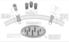
          Introduction: Systemic lupus erythematosus [SLE] is a chronic, autoimmune condition characterized by the formation of autoantibodies directed against nuclear components and by oxidative stress. Recently, a number of studies have demonstrated the essential role of iron in the immune response and there is growing evidence that abnormal iron homeostasis can occur in the chronic inflammatory state seen in SLE. Not only is iron vital for hematopoiesis, it is also important for a number of other key physiological processes, in particular in maintaining healthy mitochondrial function.

          Areas covered: In this review, we highlight the latest understanding with regards to how patients with SLE may be at risk of cellular iron depletion as a result of both absolute and functional iron deficiency. Furthermore, we aim to explain the latest evidence of mitochondrial dysfunction in the pathogenesis of the disease.

          Expert opinion: Growing evidence suggests that both abnormal iron homeostasis and subsequent mitochondrial dysfunction can impair effector immune cell function. Through a greater understanding of these abnormalities, therapeutic options that directly target iron and mitochondria may ultimately represent novel treatment targets that may translate into clinical care of patients with SLE in the near future.

          Related collections

          Most cited references105

          • Record: found
          • Abstract: found
          • Article: not found

          Ferroptosis: an iron-dependent form of nonapoptotic cell death.

          Nonapoptotic forms of cell death may facilitate the selective elimination of some tumor cells or be activated in specific pathological states. The oncogenic RAS-selective lethal small molecule erastin triggers a unique iron-dependent form of nonapoptotic cell death that we term ferroptosis. Ferroptosis is dependent upon intracellular iron, but not other metals, and is morphologically, biochemically, and genetically distinct from apoptosis, necrosis, and autophagy. We identify the small molecule ferrostatin-1 as a potent inhibitor of ferroptosis in cancer cells and glutamate-induced cell death in organotypic rat brain slices, suggesting similarities between these two processes. Indeed, erastin, like glutamate, inhibits cystine uptake by the cystine/glutamate antiporter (system x(c)(-)), creating a void in the antioxidant defenses of the cell and ultimately leading to iron-dependent, oxidative death. Thus, activation of ferroptosis results in the nonapoptotic destruction of certain cancer cells, whereas inhibition of this process may protect organisms from neurodegeneration. Copyright © 2012 Elsevier Inc. All rights reserved.
            Bookmark
            • Record: found
            • Abstract: found
            • Article: found
            Is Open Access

            Ferroptosis: past, present and future

            Ferroptosis is a new type of cell death that was discovered in recent years and is usually accompanied by a large amount of iron accumulation and lipid peroxidation during the cell death process; the occurrence of ferroptosis is iron-dependent. Ferroptosis-inducing factors can directly or indirectly affect glutathione peroxidase through different pathways, resulting in a decrease in antioxidant capacity and accumulation of lipid reactive oxygen species (ROS) in cells, ultimately leading to oxidative cell death. Recent studies have shown that ferroptosis is closely related to the pathophysiological processes of many diseases, such as tumors, nervous system diseases, ischemia-reperfusion injury, kidney injury, and blood diseases. How to intervene in the occurrence and development of related diseases by regulating cell ferroptosis has become a hotspot and focus of etiological research and treatment, but the functional changes and specific molecular mechanisms of ferroptosis still need to be further explored. This paper systematically summarizes the latest progress in ferroptosis research, with a focus on providing references for further understanding of its pathogenesis and for proposing new targets for the treatment of related diseases.
              Bookmark
              • Record: found
              • Abstract: found
              • Article: not found

              Mitochondrial energetics in the kidney

              Mitochondria provide the kidney with energy to remove waste from the blood and regulate fluid and electrolyte balance. This Review discusses how mitochondrial homeostasis is maintained, the changes in mitochondrial energetics that occur in acute kidney injury and diabetic nephropathy, and how targeting mitochondrial energetics might aid the treatment of renal disease.
                Bookmark

                Author and article information

                Journal
                Expert Rev Clin Immunol
                Expert Rev Clin Immunol
                Expert Review of Clinical Immunology
                Taylor & Francis
                1744-666X
                1744-8409
                30 July 2021
                2021
                30 July 2021
                : 17
                : 9
                : 957-967
                Affiliations
                [0001]Department of Rheumatology, Division of Medicine, University College London; , London, UK
                Author notes
                CONTACT Chris Wincup anisur.rahman@ 123456ucl.ac.uk Department of Rheumatology, Division of Medicine, University College London; , London, UK
                Article
                1953981
                10.1080/1744666X.2021.1953981
                8452144
                34263712
                d1ed5e87-e697-4055-b92b-babf629f107f
                © 2021 The Author(s). Published by Informa UK Limited, trading as Taylor & Francis Group.

                This is an Open Access article distributed under the terms of the Creative Commons Attribution-NonCommercial-NoDerivatives License ( http://creativecommons.org/licenses/by-nc-nd/4.0/), which permits non-commercial re-use, distribution, and reproduction in any medium, provided the original work is properly cited, and is not altered, transformed, or built upon in any way.

                History
                Page count
                Figures: 3, References: 105, Pages: 12
                Categories
                Research Article
                Review

                ferroptosis,functional iron deficiency,immunometabolism,iron,mitochondria,systemic lupus erythematosus

                Comments

                Comment on this article