3,583
views
2
recommends
+1 Recommend
2 collections
    57
    shares
      scite_
       
      • Record: found
      • Abstract: found
      • Article: found
      Is Open Access

      MitraClip procedure with two MitraClips after indirect annuloplasty with the MONARC device

      Published
      Clinical Case Report
      Bookmark

            Abstract

            Mitral regurgitation is associated with a worsened prognosis in dilated cardiomyopathy. First standard therapy consists of a mitral valve reconstruction through heart surgery including the heart–lung machine. In patients with high comorbidity, catheter-based techniques have been developed. In the Evolution I study, the MONARC system was implanted in the coronary sinus in the functional mitral regurgitation. A reduction in regurgitation by >1 grade was documented in 50% of the patients. MitraClip is an alternative, edge-to-edge technique, which joined the posterior and anterior leaflet by implanting a clip. It can be used for both functional and degenerative mitral regurgitations. We reported a case of MitraClip procedure with the use of two clips and a reduction of mitral regurgitation to grade 0–1 after implanting a MONARC device four years ago with missing relevant reduction in mitral regurgitation. With this report, we illustrated the management of Mitraclip in a patient with an implanted MONARC device and technical difficulties through the bowing of the posterior annulus.

            Main article text

            INTRODUCTION

            Mitral regurgitation in dilated cardiomyopathy is often a functional reason of either ischemic or idiopathic origin. Persistent severe mitral regurgitation leads to left ventricular and atrial chamber remodeling with poor clinical outcomes [1]. In most patients, effective surgical repair of the mitral valve causes a decrease in the left ventricular volume overload and leads to reverse left ventricular and left atrial remodeling [2]. In cases of patients with high comorbidity and higher mortality after surgery, less invasive catheter-based techniques have been developed. The MONARC device (Edwards Livesciences) is implanted in the coronary sinus as an indirect annuloplasty. It is constructed of nitinol and consists of a larger proximal anchor, a bridge segment with bio-absorbing coating, and a smaller distal anchor. In the Evolution I study, a patient with functional mitral regurgitation between 2+ and 4+ revealed at 12 months a reduction by >1 grade in 50%, that is less than what it obtained with surgical mitral valve repair. Patients with severe mitral regurgitation (grade >3+) seemed most likely benefitted [3]. Mitral valve repair is performed to reduce the mitral regurgitation by creating a double orifice that was first performed by Alfiri [4]. Based on this surgical technique, endovascular mitral repair has been developed with the use of a clip [5]. The posterior and the anterior leaflets are joined by implanting one or more clips resulting in reduction of mitral regurgitation. Percutaneous mitral valve repair in edge-to-edge procedure with the MitalClip device has been shown to be associated with good clinical outcomes in patients with high risk for surgery [6]. Furthermore, it demonstrated superior safety compared to surgical mitral valve repair with similar improvement in clinical outcomes [7]. In contrast to the MONARC device, the MitraClip can be used for both functional and structural mitral regurgitations.

            CASE REPORT

            We describe a case of a 73-year-old man with severe mitral regurgitation in ischemic cardiomyopathy and implanted MONARC device four years ago. The medical history included an ischemic cardiomyopathy with highly reduced ejection fraction (30%). He had a single bypass surgery in 1995 (RIVA) and stenting of the RCX in 2009. A defibrillator was implanted in case of ventricular tachycardia. Furthermore, he had impaired renal function and a moderately combined lung disease (Euroscore 51%). In 2009, he was included in the Evolutian trial and got uncomplicated a 120 cm 6/15 mm anchor (Figure 1) with a reduction of the mitral regurgitation from grade 3 to 2 after six months. Since the end of 2010, a continuing increase in mitral regurgitation was documented with a parallel increase in the New York Heart Association (NYHA) class. In the beginning of 2013 he had dyspnoea with minimal exercise and a documented high grade mitral regurgitation. So we decided on the implantation of a MitraClip.

            Figure 1.
            MONARC device implantation in 2009.

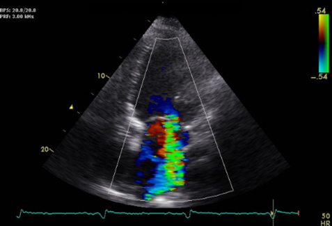
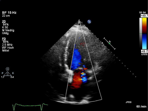
            The pre-procedural echocardiogram revealed a dilated left ventricle with end-diastolic dimension of 71 mm and serve reduced left ventricular function with 35%. We had a bi-atrial dilatation with 213 ml for the left atrium and 112 ml for the right atrium with severe mitral regurgitation and minimal tricuspid regurgitation (Figure 2). The right ventricular systolic pressure was 45 mmHg, confirming pulmonary hypertension. The trans-esophageal echocardiogram conformed severe mitral regurgitation (Figure 3). With a three-dimensional echocardiogram, we evaluated the possibility of MitraClip by enough leaflet substance without presentation of serve prolapse or structural disintegrity. Through the implanted MONARC device, a bowing posterior annulus was documented and it was difficult to evaluate the posterior leaflet length and integrity (Figure 4).

            Figure 2.
            Pre-procedural echocardiogram with bi-atrial dilatation.
            Figure 3.
            Pre-procedural trans-esophageal echocardiogram with serve mitral regurgitation.
            Figure 4.
            Echocardiogram of the MONARC device with bowing the posterior annulus and difficulty to evaluate the posterior leaflet length and integrity.

            The standard procedure was performed with percutaneous femoral venous transseptal access. The clip was aligned above the mitral valve, approved with a three-dimensional echocardiogram, before grasping and coapting the leaflets. The grasp of the leaflets was difficult in case of the bowing posterior annulus and shadow of the MONARC device. After the first grasping and clip placement, we demonstrated a reduction of the mitral regurgitation from grade 3 to 1–2 with a remaining jet lateral of the clip. The verification of the leaflet insertion confirmed enough stuff of the posterior leaflet as well as of the anterior leaflet in the clip. With the second clip, we could reduce the mitral regurgitation to grade 0–1 (Figure 5, Supplementary Video 1).

            Figure 5.
            Echocardiogram on day 3, post-procedural with residual mitral regurgitation.
            Supplementary Video 1.
            Trans-esophageal three chamber view with residual mitral regurgitation after two mitraclips.

            The procedure was completed and the anesthesia was stopped. The patient was awake and hemodynamically steady. The post-interventional echocardiogram on day 3 confirmed minimal residual regurgitation without stenosis and a re-gradient right ventricular systolic pressure to 32 mmHg by constant minimal tricuspid regurgitation.

            DISCUSSION

            The MONARC device implantation showed a response of 50% with the reduction in mitral regurgitation of grade 1 after 6–12 months, and the patients with severe initial mitral regurgitation seemed most likely benefitted. Furthermore, only the functional regurgitation is influenced. Mitral clipping is an attractive alternative treatment of severe mitral regurgitation, and, in contrast, it can be used for functional and structural mitral valve regurgitations. In addition, we have a direct effect of reduction after clipping, and the clinical improvement is instant.

            References

            1. , , , , . Relation of frequency and severity of mitral regurgitation to survival among patients with left ventricular systolic dysfunction and heart failure. Am J Cardiol. 2003;91(5):538–43. [Cross Ref]

            2. , , , , , , , . Recurrence of mitral regurgitation parallels the absence of left ventricular reverse remodeling after mitral repair in advanced dilated cardiomyopathy. Ann Thorac Surg. 2008;85(3):932–39. [Cross Ref]

            3. , , , , , , , , . Transcatheter implantation of the MONARC coronary sinus devise for mitral regurgitation: 1-year results from the EVOLUTION phase I study. J Am Coll Cardiol. 2011;4(1):115–122. [Cross Ref]

            4. , , , , , , . The double-orifice technique in mitral valve repair: a simple solution for complex problems. J Thorac Cardiovasc Surg. 2001;122(4):674–81. [Cross Ref]

            5. , , , , , , , . Endovascular edge-to-edge mitral valve repair: short-term results in a porcine model. Circulation. 2003;108(16):1990–93. [Cross Ref]

            6. , , , , , , , , , , , , , , , , , . Acute and 12 month results with catheter-based mitral valve leaflet repair: the EVEREST II (Endovascular Valve Edge to Edge Repair) high risk study. J Am Coll Cardiol. 2012;59(2):130–39. [Cross Ref]

            7. , , , , , , , , , , , , , , , , . EVEREST II Investigators. Percutaneous repair or surgery for mitral regurgitation. N Engl J Med. 2011;364(15):1395–406. [Cross Ref]

            Competing Interests

            The authors declare no competing interests.

            Publishing Notes

            © 2014 S. Rutschow et al. This work has been published open access under Creative Commons Attribution License CC BY 4.0, which permits unrestricted use, distribution, and reproduction in any medium, provided the original work is properly cited. Conditions, terms of use and publishing policy can be found at www.scienceopen.com.

            Author and article information

            Contributors
            (View ORCID Profile)
            Journal
            SOR-MED
            ScienceOpen Research
            ScienceOpen
            2199-1006
            20 October 2014
            : 0 (ID: d0c9107d-0684-4c10-b620-eca9ef5a0600 )
            : 0
            : 1-3
            Affiliations
            [0001]Department of Cardiology and Pulmology, Charite, Campus Benjamin Franklin, Hindenburgdamm 30, 12200 Berlin, Germany
            Author notes
            [* ]Corresponding author's e-mail address: susanne.rutschow@ 123456charite.de
            Article
            1953:XE
            10.14293/S2199-1006.1.SOR-MED.AO7DCV.v1
            d0c9107d-0684-4c10-b620-eca9ef5a0600
            © 2014 S. Rutschow et al.

            This work has been published open access under Creative Commons Attribution License CC BY 4.0 , which permits unrestricted use, distribution, and reproduction in any medium, provided the original work is properly cited. Conditions, terms of use and publishing policy can be found at www.scienceopen.com .

            History
            Page count
            Figures: 5, Tables: 0, References: 7, Pages: 3
            Categories
            Clinical Case Report

            Medicine
            ischaemic cardiomyopathy,MONARC device,MitraClip

            Comments

            In the manuscript “MitraClip procedure with two MitraClips after indirect annuloplasty with the MONARC device” dr. Rutschow and colleagues report on a case of severe MR in the context of LV cardiomyopathy treated with Mitraclip. The patient had undergone a Monarc device implantation four years before, but MR reduction had been incomplete and transient.

            The case is interesting as the authors address the crucial topic of high risk patients requiring treatment of severe MR. However it shows some limitations that need to be addressed in order to improve the manuscript.

            Major comments.

            The authors should concisely explain the reason for residual MR and its worsening in a patient with a Monarc device.

            Also they should answer to some questions.

            The screening TEE exam seems quite poor. No data are reported on MV leaflets characteristics and length, and no data are available of annular dimensions. A Mitraclip device is only 16 mm long when its arms are open, therefore in dilated ventricles extreme dilation of MV annulus has to be taken into account as a limiting factor. How long was the AP MV diameter in this case? What about the tethering? Was it symmetric? How deep was the coaptation point?

            Concerning the post grasping TEE, what about the residual MV area? How was this assessed?

            Considering the effect of general anaesthesia on hemodynamics and on MR, which criteria did the authors use to assess residual MR under general anaesthesia and mechanical ventilation?

            Is an immediate good reduction in MR a guarantee of competent MV on the long distance following Mitraclip?

            Minor comment

            The English language needs to be revised and several typos need to be amended. The supplemental material does not work.

            2015-05-08 14:48 UTC
            +1
            I am happy to review this interesting paper regarding the use of MitraClip for challenging cases. MitraClip has gained wide clinical since it can be used for treating both functional and degenerative mitral regurgitation. Moreover, recent publications have addressed that this device may be useful as well in different and complex clinical scenarios such as acute MR after AMI, failed annuloplasty rings or MR associated to hypertrophic cardiomyopathy. This interesting paper highlights a new use for the MitraClip. Two clips were implanted in a patient with a prior indirect annuloplasty device and nearly complete reduction of MR was achieved. The case is appropriate and the images are interesting. However, the paper has as well some shortcomings. The English style is far from ideal and some sentences are poorly built. There is no description of how operators dealt with this difficult PML or if they used a special image technique for increasing the visibility of such leaflet.
            2014-12-24 11:21 UTC
            +1
            2 people recommend this

            Comment on this article