51
views
0
recommends
+1 Recommend
0 collections
    0
    shares
      • Record: found
      • Abstract: found
      • Article: found
      Is Open Access

      Development of structure–function coupling in human brain networks during youth

      research-article

      Read this article at

      Bookmark
          There is no author summary for this article yet. Authors can add summaries to their articles on ScienceOpen to make them more accessible to a non-specialist audience.

          Significance

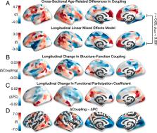
          The human brain is organized into a hierarchy of functional systems that evolve in childhood and adolescence to support the dynamic control of attention and behavior. However, it remains unknown how developing white-matter architecture supports coordinated fluctuations in neural activity underlying cognition. We document marked remodeling of structure–function coupling in youth, which aligns with cortical hierarchies of functional specialization and evolutionary expansion. Further, we demonstrate that structure–function coupling in rostrolateral prefrontal cortex supports age-related improvements in executive ability. These findings have broad relevance for accounts of experience-dependent plasticity in healthy development and abnormal development associated with neuropsychiatric illness.

          Abstract

          The protracted development of structural and functional brain connectivity within distributed association networks coincides with improvements in higher-order cognitive processes such as executive function. However, it remains unclear how white-matter architecture develops during youth to directly support coordinated neural activity. Here, we characterize the development of structure–function coupling using diffusion-weighted imaging and n-back functional MRI data in a sample of 727 individuals (ages 8 to 23 y). We found that spatial variability in structure–function coupling aligned with cortical hierarchies of functional specialization and evolutionary expansion. Furthermore, hierarchy-dependent age effects on structure–function coupling localized to transmodal cortex in both cross-sectional data and a subset of participants with longitudinal data ( n = 294). Moreover, structure–function coupling in rostrolateral prefrontal cortex was associated with executive performance and partially mediated age-related improvements in executive function. Together, these findings delineate a critical dimension of adolescent brain development, whereby the coupling between structural and functional connectivity remodels to support functional specialization and cognition.

          Related collections

          Most cited references29

          • Record: found
          • Abstract: found
          • Article: not found

          Situating the default-mode network along a principal gradient of macroscale cortical organization.

          Understanding how the structure of cognition arises from the topographical organization of the cortex is a primary goal in neuroscience. Previous work has described local functional gradients extending from perceptual and motor regions to cortical areas representing more abstract functions, but an overarching framework for the association between structure and function is still lacking. Here, we show that the principal gradient revealed by the decomposition of connectivity data in humans and the macaque monkey is anchored by, at one end, regions serving primary sensory/motor functions and at the other end, transmodal regions that, in humans, are known as the default-mode network (DMN). These DMN regions exhibit the greatest geodesic distance along the cortical surface-and are precisely equidistant-from primary sensory/motor morphological landmarks. The principal gradient also provides an organizing spatial framework for multiple large-scale networks and characterizes a spectrum from unimodal to heteromodal activity in a functional metaanalysis. Together, these observations provide a characterization of the topographical organization of cortex and indicate that the role of the DMN in cognition might arise from its position at one extreme of a hierarchy, allowing it to process transmodal information that is unrelated to immediate sensory input.
            Bookmark
            • Record: found
            • Abstract: found
            • Article: not found

            The evolution of distributed association networks in the human brain.

            The human cerebral cortex is vastly expanded relative to other primates and disproportionately occupied by distributed association regions. Here we offer a hypothesis about how association networks evolved their prominence and came to possess circuit properties vital to human cognition. The rapid expansion of the cortical mantle may have untethered large portions of the cortex from strong constraints of molecular gradients and early activity cascades that lead to sensory hierarchies. What fill the gaps between these hierarchies are densely interconnected networks that widely span the cortex and mature late into development. Limitations of the tethering hypothesis are discussed as well as its broad implications for understanding critical features of the human brain as a byproduct of size scaling. Copyright © 2013 Elsevier Ltd. All rights reserved.
              Bookmark
              • Record: found
              • Abstract: not found
              • Article: not found

              The anatomical basis of functional localization in the cortex.

                Bookmark

                Author and article information

                Journal
                Proc Natl Acad Sci U S A
                Proc. Natl. Acad. Sci. U.S.A
                pnas
                pnas
                PNAS
                Proceedings of the National Academy of Sciences of the United States of America
                National Academy of Sciences
                0027-8424
                1091-6490
                7 January 2020
                24 December 2019
                24 December 2019
                : 117
                : 1
                : 771-778
                Affiliations
                [1] aDepartment of Psychiatry, University of Pennsylvania , Philadelphia, PA 19104;
                [2] bLifespan Brain Institute, Children’s Hospital of Philadelphia , Philadelphia, PA 19104;
                [3] cDepartment of Bioengineering, Stanford University , Stanford, CA 94305;
                [4] dDepartment of Psychological and Brain Sciences, Indiana University Bloomington , Bloomington, IN 47405;
                [5] eDepartment of Radiology, University of Pennsylvania , Philadelphia, PA 19104;
                [6] fDepartment of Psychiatry, Yale University School of Medicine , New Haven, CT 06510;
                [7] gPenn Statistics in Imaging and Visualization Center, Department of Biostatistics, Epidemiology and Informatics, University of Pennsylvania , Philadelphia, PA 19104;
                [8] hCenter for Biomedical Image Computing and Analytics, University of Pennsylvania , Philadelphia, PA 19104;
                [9] iDevelopmental Neurogenomics Unit, National Institute of Mental Health , Bethesda, MD 20814;
                [10] jDepartment of Neurology, University of Pennsylvania , Philadelphia, PA 19104;
                [11] kDepartment of Bioengineering, University of Pennsylvania , Philadelphia, PA 19104;
                [12] lDepartment of Electrical and Systems Engineering, University of Pennsylvania , Philadelphia, PA 19104;
                [13] mDepartment of Physics and Astronomy, University of Pennsylvania , Philadelphia, PA 19104;
                [14] nSanta Fe Institute , Santa Fe, NM 87501
                Author notes
                1To whom correspondence may be addressed. Email: sattertt@ 123456pennmedicine.upenn.edu .

                Edited by Marcus E. Raichle, Washington University in St. Louis, St. Louis, MO, and approved November 27, 2019 (received for review July 12, 2019)

                Author contributions: G.L.B., R.E.G., R.C.G., D.S.B., and T.D.S. designed research; G.L.B. performed research; Z.C., D.R.R., R.C., R.F.B., B.L., M.C., P.A.C., C.H.X., T.M.M., K.R., D.J.O., A.F.A.-B., R.T.S., A.R., D.S.B., and T.D.S. contributed new reagents/analytic tools; G.L.B., Z.C., R.C., and T.M.M. analyzed data; and G.L.B. and T.D.S. wrote the paper.

                Author information
                http://orcid.org/0000-0001-6554-1893
                http://orcid.org/0000-0001-8627-8203
                http://orcid.org/0000-0002-6183-4493
                http://orcid.org/0000-0001-7072-9399
                Article
                201912034
                10.1073/pnas.1912034117
                6955327
                31874926
                d0b93702-fd70-4b72-adb2-71ad97f05673
                Copyright © 2020 the Author(s). Published by PNAS.

                This open access article is distributed under Creative Commons Attribution-NonCommercial-NoDerivatives License 4.0 (CC BY-NC-ND).

                History
                Page count
                Pages: 8
                Funding
                Funded by: HHS | NIH | National Institute of Mental Health (NIMH) 100000025
                Award ID: F31MH115709
                Award Recipient : Graham L. Baum Award Recipient : Theodore D. Satterthwaite
                Funded by: HHS | NIH | National Institute of Mental Health (NIMH) 100000025
                Award ID: R01MH113550
                Award Recipient : Graham L. Baum Award Recipient : Theodore D. Satterthwaite
                Categories
                Biological Sciences
                Psychological and Cognitive Sciences

                brain development,mri,connectome,cortical organization,structure–function

                Comments

                Comment on this article