16
views
0
recommends
+1 Recommend
0 collections
    0
    shares
      • Record: found
      • Abstract: found
      • Article: found
      Is Open Access

      There is more than one way to turn a spherical cellular monolayer inside out: type B embryo inversion in Volvox globator

      research-article
      1 , 1 ,
      BMC Biology
      BioMed Central

      Read this article at

      Bookmark
          There is no author summary for this article yet. Authors can add summaries to their articles on ScienceOpen to make them more accessible to a non-specialist audience.

          Abstract

          Background

          Epithelial folding is a common morphogenetic process during the development of multicellular organisms. In metazoans, the biological and biomechanical processes that underlie such three-dimensional (3D) developmental events are usually complex and difficult to investigate. Spheroidal green algae of the genus Volvox are uniquely suited as model systems for studying the basic principles of epithelial folding. Volvox embryos begin life inside out and then must turn their spherical cell monolayer outside in to achieve their adult configuration; this process is called 'inversion.' There are two fundamentally different sequences of inversion processes in Volvocaceae: type A and type B. Type A inversion is well studied, but not much is known about type B inversion. How does the embryo of a typical type B inverter, V. globator, turn itself inside out?

          Results

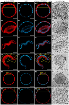
          In this study, we investigated the type B inversion of V. globator embryos and focused on the major movement patterns of the cellular monolayer, cell shape changes and changes in the localization of cytoplasmic bridges (CBs) connecting the cells. Isolated intact, sectioned and fragmented embryos were analyzed throughout the inversion process using light microscopy, confocal laser scanning microscopy, scanning electron microscopy and transmission electron microscopy techniques. We generated 3D models of the identified cell shapes, including the localizations of CBs. We show how concerted cell-shape changes and concerted changes in the position of cells relative to the CB system cause cell layer movements and turn the spherical cell monolayer inside out. The type B inversion of V. globator is compared to the type A inversion in V. carteri.

          Conclusions

          Concerted, spatially and temporally coordinated changes in cellular shapes in conjunction with concerted migration of cells relative to the CB system are the causes of type B inversion in V. globator. Despite significant similarities between type A and type B inverters, differences exist in almost all details of the inversion process, suggesting analogous inversion processes that arose through parallel evolution. Based on our results and due to the cellular biomechanical implications of the involved tensile and compressive forces, we developed a global mechanistic scenario that predicts epithelial folding during embryonic inversion in V. globator.

          Related collections

          Most cited references68

          • Record: found
          • Abstract: found
          • Article: not found

          Tissue tectonics: morphogenetic strain rates, cell shape change and intercalation

          The dynamic reshaping of tissues during morphogenesis results from a combination of individual cell behaviours and collective cell rearrangements. However, a comprehensive framework to unambiguously measure and link cell behaviour to tissue morphogenesis is lacking. Here we introduce such a kinematic framework, bridging cell and tissue behaviours at an intermediate, mesoscopic, level of cell clusters or domains. By measuring domain deformation in terms of the relative motion of cell positions and the evolution of their shapes, we characterize the basic invariant quantities that measure fundamental classes of cell behaviour, namely tensorial rates of cell shape change and cell intercalation. In doing so we introduce an explicit definition of cell intercalation as a continuous process. We demonstrate how spatiotemporal mapping of strain rates in three models of tissue morphogenesis leads to new insight into morphogenetic mechanisms. Our quantitative approach has broad relevance for the precise characterisation and comparison of morphogenetic phenotypes.
            Bookmark
            • Record: found
            • Abstract: found
            • Article: not found

            How we are shaped: the biomechanics of gastrulation.

            Although it is rarely considered so in modern developmental biology, morphogenesis is fundamentally a biomechanical process, and this is especially true of one of the first major morphogenic transformations in development, gastrulation. Cells bring about changes in embryonic form by generating patterned forces and by differentiating the tissue mechanical properties that harness these forces in specific ways. Therefore, biomechanics lies at the core of connecting the genetic and molecular basis of cell activities to the macroscopic tissue deformations that shape the embryo. Here we discuss what is known of the biomechanics of gastrulation, primarily in amphibians but also comparing similar morphogenic processes in teleost fish and amniotes, and selected events in several species invertebrates. Our goal is to review what is known and identify problems for further research.
              Bookmark
              • Record: found
              • Abstract: found
              • Article: not found

              Cell shape changes indicate a role for extrinsic tensile forces in Drosophila germ-band extension.

              Drosophila germ-band extension (GBE) is an example of the convergence and extension movements that elongate and narrow embryonic tissues. To understand the collective cell behaviours underlying tissue morphogenesis, we have continuously quantified cell intercalation and cell shape change during GBE. We show that the fast, early phase of GBE depends on cell shape change in addition to cell intercalation. In antero-posterior patterning mutants such as those for the gap gene Krüppel, defective polarized cell intercalation is compensated for by an increase in antero-posterior cell elongation, such that the initial rate of extension remains the same. Spatio-temporal patterns of cell behaviours indicate that an antero-posterior tensile force deforms the germ band, causing the cells to change shape passively. The rate of antero-posterior cell elongation is reduced in twist mutant embryos, which lack mesoderm. We propose that cell shape change contributing to germ-band extension is a passive response to mechanical forces caused by the invaginating mesoderm.
                Bookmark

                Author and article information

                Journal
                BMC Biol
                BMC Biol
                BMC Biology
                BioMed Central
                1741-7007
                2011
                29 December 2011
                : 9
                : 89
                Affiliations
                [1 ]Department of Cellular and Developmental Biology of Plants, University of Bielefeld, Universitätsstrasse 25, 33615 Bielefeld, Germany
                Article
                1741-7007-9-89
                10.1186/1741-7007-9-89
                3324393
                22206406
                d016939e-16a9-416a-9e7a-12c80c8cbec9
                Copyright ©2011 Höhn and Hallmann; licensee BioMed Central Ltd.

                This is an Open Access article distributed under the terms of the Creative Commons Attribution License ( http://creativecommons.org/licenses/by/2.0), which permits unrestricted use, distribution, and reproduction in any medium, provided the original work is properly cited.

                History
                : 25 August 2011
                : 29 December 2011
                Categories
                Research Article

                Life sciences
                Life sciences

                Comments

                Comment on this article