67
views
0
recommends
+1 Recommend
0 collections
    0
    shares
      • Record: found
      • Abstract: found
      • Article: found
      Is Open Access

      Wallerian degeneration: gaining perspective on inflammatory events after peripheral nerve injury

      review-article

      Read this article at

      Bookmark
          There is no author summary for this article yet. Authors can add summaries to their articles on ScienceOpen to make them more accessible to a non-specialist audience.

          Abstract

          In this review, we first provide a brief historical perspective, discussing how peripheral nerve injury (PNI) may have caused World War I. We then consider the initiation, progression, and resolution of the cellular inflammatory response after PNI, before comparing the PNI inflammatory response with that induced by spinal cord injury (SCI).

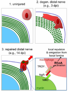
          In contrast with central nervous system (CNS) axons, those in the periphery have the remarkable ability to regenerate after injury. Nevertheless, peripheral nervous system (PNS) axon regrowth is hampered by nerve gaps created by injury. In addition, the growth-supportive milieu of PNS axons is not sustained over time, precluding long-distance regeneration. Therefore, studying PNI could be instructive for both improving PNS regeneration and recovery after CNS injury. In addition to requiring a robust regenerative response from the injured neuron itself, successful axon regeneration is dependent on the coordinated efforts of non-neuronal cells which release extracellular matrix molecules, cytokines, and growth factors that support axon regrowth. The inflammatory response is initiated by axonal disintegration in the distal nerve stump: this causes blood-nerve barrier permeabilization and activates nearby Schwann cells and resident macrophages via receptors sensitive to tissue damage. Denervated Schwann cells respond to injury by shedding myelin, proliferating, phagocytosing debris, and releasing cytokines that recruit blood-borne monocytes/macrophages. Macrophages take over the bulk of phagocytosis within days of PNI, before exiting the nerve by the circulation once remyelination has occurred. The efficacy of the PNS inflammatory response (although transient) stands in stark contrast with that of the CNS, where the response of nearby cells is associated with inhibitory scar formation, quiescence, and degeneration/apoptosis. Rather than efficiently removing debris before resolving the inflammatory response as in other tissues, macrophages infiltrating the CNS exacerbate cell death and damage by releasing toxic pro-inflammatory mediators over an extended period of time. Future research will help determine how to manipulate PNS and CNS inflammatory responses in order to improve tissue repair and functional recovery.

          Related collections

          Most cited references136

          • Record: found
          • Abstract: found
          • Article: not found

          Two types of murine helper T cell clone. I. Definition according to profiles of lymphokine activities and secreted proteins.

          A panel of antigen-specific mouse helper T cell clones was characterized according to patterns of lymphokine activity production, and two types of T cell were distinguished. Type 1 T helper cells (TH1) produced IL 2, interferon-gamma, GM-CSF, and IL 3 in response to antigen + presenting cells or to Con A, whereas type 2 helper T cells (TH2) produced IL 3, BSF1, and two other activities unique to the TH2 subset, a mast cell growth factor distinct from IL 3 and a T cell growth factor distinct from IL 2. Clones representing each type of T cell were characterized, and the pattern of lymphokine activities was consistent within each set. The secreted proteins induced by Con A were analyzed by biosynthetic labeling and SDS gel electrophoresis, and significant differences were seen between the two groups of T cell line. Both types of T cell grew in response to alternating cycles of antigen stimulation, followed by growth in IL 2-containing medium. Examples of both types of T cell were also specific for or restricted by the I region of the MHC, and the surface marker phenotype of the majority of both types was Ly-1+, Lyt-2-, L3T4+, Both types of helper T cell could provide help for B cells, but the nature of the help differed. TH1 cells were found among examples of T cell clones specific for chicken RBC and mouse alloantigens. TH2 cells were found among clones specific for mouse alloantigens, fowl gamma-globulin, and KLH. The relationship between these two types of T cells and previously described subsets of T helper cells is discussed.
            Bookmark
            • Record: found
            • Abstract: found
            • Article: not found

            Identification of the Nogo inhibitor of axon regeneration as a Reticulon protein.

            Adult mammalian axon regeneration is generally successful in the peripheral nervous system (PNS) but is dismally poor in the central nervous system (CNS). However, many classes of CNS axons can extend for long distances in peripheral nerve grafts. A comparison of myelin from the CNS and the PNS has revealed that CNS white matter is selectively inhibitory for axonal outgrowth. Several components of CNS white matter, NI35, NI250(Nogo) and MAG, that have inhibitory activity for axon extension have been described. The IN-1 antibody, which recognizes NI35 and NI250(Nogo), allows moderate degrees of axonal regeneration and functional recovery after spinal cord injury. Here we identify Nogo as a member of the Reticulon family, Reticulon 4-A. Nogo is expressed by oligodendrocytes but not by Schwann cells, and associates primarily with the endoplasmic reticulum. A 66-residue lumenal/extracellular domain inhibits axonal extension and collapses dorsal root ganglion growth cones. In contrast to Nogo, Reticulon 1 and 3 are not expressed by oligodendrocytes, and the 66-residue lumenal/extracellular domains from Reticulon 1, 2 and 3 do not inhibit axonal regeneration. These data provide a molecular basis to assess the contribution of Nogo to the failure of axonal regeneration in the adult CNS.
              Bookmark
              • Record: found
              • Abstract: found
              • Article: not found

              Wallerian degeneration, wld(s), and nmnat.

              Traditionally, researchers have believed that axons are highly dependent on their cell bodies for long-term survival. However, recent studies point to the existence of axon-autonomous mechanism(s) that regulate rapid axon degeneration after axotomy. Here, we review the cellular and molecular events that underlie this process, termed Wallerian degeneration. We describe the biphasic nature of axon degeneration after axotomy and our current understanding of how Wld(S)--an extraordinary protein formed by fusing a Ube4b sequence to Nmnat1--acts to protect severed axons. Interestingly, the neuroprotective effects of Wld(S) span all species tested, which suggests that there is an ancient, Wld(S)-sensitive axon destruction program. Recent studies with Wld(S) also reveal that Wallerian degeneration is genetically related to several dying back axonopathies, thus arguing that Wallerian degeneration can serve as a useful model to understand, and potentially treat, axon degeneration in diverse traumatic or disease contexts.
                Bookmark

                Author and article information

                Journal
                J Neuroinflammation
                Journal of Neuroinflammation
                BioMed Central
                1742-2094
                2011
                30 August 2011
                : 8
                : 110
                Affiliations
                [1 ]Department of Neuroscience and Center for Brain and Spinal Cord Repair, College of Medicine, The Ohio State University, 770 Biomedical Research Tower, 460 West 12th Ave, Columbus, OH, 43210, USA
                [2 ]International Collaboration On Repair Discoveries (ICORD), Vancouver Coastal Health Research Institute, and Department of Zoology, University of British Columbia, 818 West 10th Ave, Vancouver, BC, V5T 1M9, Canada
                Article
                1742-2094-8-110
                10.1186/1742-2094-8-110
                3180276
                21878126
                cf13d3fa-ce71-4778-8acb-9881ecb8dc85
                Copyright ©2011 Gaudet et al; licensee BioMed Central Ltd.

                This is an Open Access article distributed under the terms of the Creative Commons Attribution License ( http://creativecommons.org/licenses/by/2.0), which permits unrestricted use, distribution, and reproduction in any medium, provided the original work is properly cited.

                History
                : 14 July 2011
                : 30 August 2011
                Categories
                Review

                Neurosciences
                inflammation,phagocytosis,axotomy,neuroinflammation,spinal cord injury,microglia,wallerian degeneration,macrophage,galectin-1

                Comments

                Comment on this article