4
views
0
recommends
+1 Recommend
0 collections
    0
    shares
      • Record: found
      • Abstract: found
      • Article: found
      Is Open Access

      Pure Tethered Cervical Cord and Review of Literature

      case-report

      Read this article at

      Bookmark
          There is no author summary for this article yet. Authors can add summaries to their articles on ScienceOpen to make them more accessible to a non-specialist audience.

          Abstract

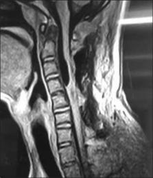
          Tethering of the spinal cord in the lumbosacral region with myelomeningocele is a well-known phenomenon. Only sporadic cases of tethering along the rest of the neuraxis, including the hindbrain, cervical, and thoracic spinal cord have been documented, always along with some associated congenital malformations (hydrocephalus, Chiari malformation, myelomeningocele, meningocele, hamartomatous stalk, spina bifida occulta, intramedullary lipoma, intradural fibrous adhesions, the fusion of the sixth and seventh cervical vertebrae, split cord malformation, or low-lying cord). In this report, 14-year-old male developed symptoms related to tethering of the cervical spinal cord, but without any associated congenital malformations, that is the pure tethered cervical cord. This causes his moribund status and makes the manuscript unique and contributes to the hitherto literature. The authors discuss the diagnosis, treatment, and postoperative course of this entity. The uniqueness in treatment is that we have operated the case without the help of intraoperative somatosensory evoked potentials and motor evoked potential from posterolateral approach under local anesthesia.

          Related collections

          Most cited references13

          • Record: found
          • Abstract: found
          • Article: not found

          Cervical myelomeningoceles.

          Cervical myelomeningoceles are rare dysraphic lesions. Nine cases of cervical myelomeningoceles are reported. The external features of all nine myelomeningoceles were strikingly similar: They were sturdy, tubular protuberances from the back of the infants' necks, covered at the base by full-thickness skin and covered on the dome by thick squamous epithelium. Internally, these were tethered cord lesions in which fibroneural bands or sagittal midline fibrous septa were tightly tethering the cervical spinal cord to the adjacent dural or intrasaccular soft tissues. Six of our early cases (Group 1) were initially treated with simple subcutaneous resection of the sac and ligation of the dural fistula without release of the internal tethering structures. Five of these children subsequently deteriorated 13 months to 8 years later, all with worsening hand function and spastic legs. All five were reexplored, and the tethering bands and septa were excised; all showed improvement. The other three neonates (Group 2) treated in the last 4 years underwent initial intradural exploration of the lesions; in one case, the tethering fibrous elements were only partially eliminated and the patient deteriorated 4 years later, but improved after a second operation for resection of a missed ventral fibrous septum. The other two Group 2 infants had a thorough release of the fibroneural stalks initially, and both were neurologically stable 3 years later. We recommend that cervical myelomeningoceles should be studied preoperatively with magnetic resonance imaging and computed tomographic myelography to identify the internal structures. The minimum initial surgical treatment should be a two-level laminectomy, intradural exploration, and excision of all tethering bands and septa, in addition to resection of the sac. If a split cord is revealed by imaging studies, both the ventral and dorsal surfaces of the hemicords must be carefully inspected to locate the median septum.
            Bookmark
            • Record: found
            • Abstract: found
            • Article: not found

            Split cervical spinal cord malformation and vertebral dysgenesis.

            We report a case of vertebral malformation associated with diplomyelia believed to be a type II split cord malformation. Cervicothoracic level cases are exceptional. This article reports the case of an 11-year-old boy with no neurological symptoms who had not undergone surgery. The diagnosis was made during pregnancy by prenatal screening with ultrasound and MRI. Several embryological theories have been offered to provide an explanation for this syndrome. Close follow-up is mandatory. Surgery must only be considered if neurological deterioration occurs. 2009 Published by Elsevier Masson SAS.
              Bookmark
              • Record: found
              • Abstract: found
              • Article: not found

              Delayed postoperative tethering of the cervical spinal cord.

              Tethering of the spinal cord in the lumbar and sacral regions of children with congenital anomalies is a well-recognized problem; however, tethering in the cervical region has rarely been reported. A search of the literature revealed no reports of symptomatic postoperative cervical spinal cord tethering. The authors present five cases of delayed postoperative cervical spinal cord tethering and discuss the benefit of detethering in these patients. All five patients were young (16 to 42 years of age) at presentation. All had done well after an initial surgical procedure but returned between 1 and 31 years postoperatively with symptoms including severe headache, upper-extremity pain, and progressive neurological deficits. In each case, magnetic resonance imaging indicated dorsal tethering of the cord in the cervical region. Surgical exploration with microscopic sharp detethering of the cervical cord was performed on each patient with favorable results. To avoid retethering, wide Tutoplast duraplasty is recommended.
                Bookmark

                Author and article information

                Journal
                Asian J Neurosurg
                Asian J Neurosurg
                AJNS
                Asian Journal of Neurosurgery
                Medknow Publications & Media Pvt Ltd (India )
                1793-5482
                2248-9614
                Jan-Mar 2018
                : 13
                : 1
                : 72-74
                Affiliations
                [1] Consultant Neurosurgeon, Mayo Hospital, Vikas Khand I, Lucknow, Uttar Pradesh, India
                [1 ] Consultant Anasthetist, Mayo Hospital, Vikas Khand I, Lucknow, Uttar Pradesh, India
                [2 ] Consultant Radiologist, MVT Diagnostics, Indiranagar, Lucknow, Uttar Pradesh, India
                [3 ] Director And Neurosurgeon, Sahara Hospital, Lucknow, Uttar Pradesh, India
                [4 ] Consultant Urologist, Neuro Center, PSS Complex, Vishal Khand III, Gomti Nagar, Lucknow, Uttar Pradesh, India
                [5 ] Director, General Surgeon, Samarpan Hospital, Lucknow, Uttar Pradesh, India
                Author notes
                Address for correspondence: Dr. Vinod Kumar Tewari, Mayo Hospital Vikas Khand I, Lucknow - 226 010, Uttar Pradesh, India. E-mail: drvinodtewari@ 123456gmail.com
                Article
                AJNS-13-72
                10.4103/1793-5482.224834
                5820900
                29492126
                cdb8db9b-7ecc-4475-b8a1-eb789c28dc38
                Copyright: © 2018 Asian Journal of Neurosurgery

                This is an open access article distributed under the terms of the Creative Commons Attribution-NonCommercial-ShareAlike 3.0 License, which allows others to remix, tweak, and build upon the work non-commercially, as long as the author is credited and the new creations are licensed under the identical terms.

                History
                Categories
                Case Report

                Surgery
                awake detethering,pure tethered cervical cord,single breadth count
                Surgery
                awake detethering, pure tethered cervical cord, single breadth count

                Comments

                Comment on this article