24
views
0
recommends
+1 Recommend
0 collections
    0
    shares
      • Record: found
      • Abstract: found
      • Article: found
      Is Open Access

      A Review of Adaptive Optics Optical Coherence Tomography: Technical Advances, Scientific Applications, and the Future

      research-article

      Read this article at

      Bookmark
          There is no author summary for this article yet. Authors can add summaries to their articles on ScienceOpen to make them more accessible to a non-specialist audience.

          Abstract

          Purpose

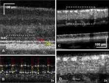
          Optical coherence tomography (OCT) has enabled “virtual biopsy” of the living human retina, revolutionizing both basic retina research and clinical practice over the past 25 years. For most of those years, in parallel, adaptive optics (AO) has been used to improve the transverse resolution of ophthalmoscopes to foster in vivo study of the retina at the microscopic level. Here, we review work done over the last 15 years to combine the microscopic transverse resolution of AO with the microscopic axial resolution of OCT, building AO-OCT systems with the highest three-dimensional resolution of any existing retinal imaging modality.

          Methods

          We surveyed the literature to identify the most influential antecedent work, important milestones in the development of AO-OCT technology, its applications that have yielded new knowledge, research areas into which it may productively expand, and nascent applications that have the potential to grow.

          Results

          Initial efforts focused on demonstrating three-dimensional resolution. Since then, many improvements have been made in resolution and speed, as well as other enhancements of acquisition and postprocessing techniques. Progress on these fronts has produced numerous discoveries about the anatomy, function, and optical properties of the retina.

          Conclusions

          Adaptive optics OCT continues to evolve technically and to contribute to our basic and clinical knowledge of the retina. Due to its capacity to reveal cellular and microscopic detail invisible to clinical OCT systems, it is an ideal companion to those instruments and has the demonstrable potential to produce images that can guide the interpretation of clinical findings.

          Related collections

          Most cited references94

          • Record: found
          • Abstract: found
          • Article: not found

          Optical coherence tomography.

          A technique called optical coherence tomography (OCT) has been developed for noninvasive cross-sectional imaging in biological systems. OCT uses low-coherence interferometry to produce a two-dimensional image of optical scattering from internal tissue microstructures in a way that is analogous to ultrasonic pulse-echo imaging. OCT has longitudinal and lateral spatial resolutions of a few micrometers and can detect reflected signals as small as approximately 10(-10) of the incident optical power. Tomographic imaging is demonstrated in vitro in the peripapillary area of the retina and in the coronary artery, two clinically relevant examples that are representative of transparent and turbid media, respectively.
            Bookmark
            • Record: found
            • Abstract: found
            • Article: not found

            Proposed lexicon for anatomic landmarks in normal posterior segment spectral-domain optical coherence tomography: the IN•OCT consensus.

            To develop a consensus nomenclature for the classification of retinal and choroidal layers and bands visible on spectral-domain optical coherence tomography (SD-OCT) images of a normal eye.
              Bookmark
              • Record: found
              • Abstract: found
              • Article: not found

              Anatomical correlates to the bands seen in the outer retina by optical coherence tomography: literature review and model.

              To evaluate the validity of commonly used anatomical designations for the four hyperreflective outer retinal bands seen in current-generation optical coherence tomography, a scale model of outer retinal morphology was created using published information for direct comparison with optical coherence tomography scans. Articles and books concerning histology of the outer retina from 1900 until 2009 were evaluated, and data were used to create a scale model drawing. Boundaries between outer retinal tissue compartments described by the model were compared with intensity variations of representative spectral-domain optical coherence tomography scans using longitudinal reflectance profiles to determine the region of origin of the hyperreflective outer retinal bands. This analysis showed a high likelihood that the spectral-domain optical coherence tomography bands attributed to the external limiting membrane (the first, innermost band) and to the retinal pigment epithelium (the fourth, outermost band) are correctly attributed. Comparative analysis showed that the second band, often attributed to the boundary between inner and outer segments of the photoreceptors, actually aligns with the ellipsoid portion of the inner segments. The third band corresponded to an ensheathment of the cone outer segments by apical processes of the retinal pigment epithelium in a structure known as the contact cylinder. Anatomical attributions and subsequent pathophysiologic assessments pertaining to the second and third outer retinal hyperreflective bands may not be correct. This analysis has identified testable hypotheses for the actual correlates of the second and third bands. Nonretinal pigment epithelium contributions to the fourth band (e.g., Bruch membrane) remain to be determined.
                Bookmark

                Author and article information

                Journal
                Invest Ophthalmol Vis Sci
                Invest. Ophthalmol. Vis. Sci
                iovs
                iovs
                iovs
                Investigative Ophthalmology & Visual Science
                The Association for Research in Vision and Ophthalmology
                0146-0404
                1552-5783
                13 July 2016
                July 2016
                : 57
                : 9
                : OCT51-OCT68
                Affiliations
                [1 ]Vision Science and Advanced Retinal Imaging Laboratory University of California-Davis, Sacramento, California, United States
                [2 ]School of Optometry, Indiana University, Bloomington, Indiana, United States
                Author notes
                Correspondence: Ravi S. Jonnal, 4860 Y Street, Suite 2400, Sacramento, CA 95817, USA; rsjonnal@ 123456ucdavis.edu .
                Article
                iovs-57-101-64 IOVS-16-19103
                10.1167/iovs.16-19103
                4968917
                27409507
                cd3a72a8-5e7e-4647-9ba3-48cfbc413cf5

                This work is licensed under a Creative Commons Attribution-NonCommercial-NoDerivatives 4.0 International License.

                History
                : 8 January 2016
                : 29 May 2016
                Categories
                Articles

                adaptive optics,optical coherence tomography,retinal imaging

                Comments

                Comment on this article