39
views
0
recommends
+1 Recommend
0 collections
    0
    shares
      • Record: found
      • Abstract: found
      • Article: found
      Is Open Access

      Stone Attenuation Value and Cross-Sectional Area on Computed Tomography Predict the Success of Shock Wave Lithotripsy

      research-article

      Read this article at

      Bookmark
          There is no author summary for this article yet. Authors can add summaries to their articles on ScienceOpen to make them more accessible to a non-specialist audience.

          Abstract

          Purpose

          To identify the parameters on noncontrast computed tomography (NCCT) that best predict the success of shock wave lithotripsy (SWL).

          Materials and Methods

          We reviewed the records of 75 patients who underwent SWL for urinary calculi measuring 5 to 20 mm. Using NCCT images, we estimated the largest stone cross-sectional area and contoured the inner edge of the stone. Clinical outcome was classified as successful (stone-free or <4 mm in diameter) or failed (stone fragments, ≥4 mm). The impact of preoperative parameters was evaluated by univariate and multivariate analysis.

          Results

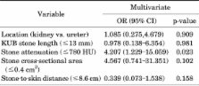
          The overall success rate was 73.3%. Average stone attenuation value, stone length, and stone cross-sectional area in the success and failure groups were 627.4±166.5 HU (Hounsfield unit) vs. 788.1±233.9 HU (p=0.002), 11.7±3.8 mm vs. 14.2±3.6 mm (p=0.015), and 0.31±0.17 cm 2 vs. 0.57±0.41 cm 2 (p<0.001), respectively. In the multivariate analysis, stone attenuation value was the only independent predictor of SWL success (p=0.023), although stone cross-sectional area had a tendency to be associated with SWL success (p=0.053). Patients were then classified into four groups by using cutoff values of 780 HU for stone attenuation value and 0.4 cm 2 for cross-sectional area. By use of these cutoff values, the group with a low stone attenuation value and a low cross-sectional area was more than 11.6 times as likely to have a successful result on SWL as were all other groups (odds ratio, 11.6; 95% confidence interval, 3.9 to 54.7; p<0.001).

          Conclusions

          Stone attenuation value and stone cross-sectional area are good predictors of extracorporeal SWL outcome.

          Related collections

          Most cited references19

          • Record: found
          • Abstract: not found
          • Article: not found

          2007 Guideline for the management of ureteral calculi.

            Bookmark
            • Record: found
            • Abstract: found
            • Article: not found

            Shock wave lithotripsy success determined by skin-to-stone distance on computed tomography.

            To evaluate whether the skin-to-stone distance (SSD), body mass index (BMI), and Hounsfield unit (HU) density can be used as independent predictors of stone-free (SF) status after shock wave lithotripsy (SWL) of lower pole kidney stones. No studies have evaluated the SSD by non-contrast-enhanced computed tomography (NCCT) as a predictor of SWL success. Studies have suggested that the BMI and HU density of urinary calculi on NCCT may predict the SF rate after SWL. The radiographs of 64 patients treated with SWL (DoliS lithotripter) from March 2000 to April 2004 with lower pole kidney stones measuring 0.5 to 1.5 cm on NCCT were reviewed. The average SSD was calculated by measuring three distances from the center of the stone to the skin (0 degrees, 45 degrees, and 90 degrees angles) on NCCT. The BMI and HU density were determined, and chemical analysis was performed on all stones. Radiographic assessment of the kidneys, ureter, and bladder at 6 weeks categorized patients into the SF or residual stone group. Logistic regression was fit, using SSD, BMI, and HU density as predictors, to assess the SF rates after SWL. Of 64 patients, 30 were SF and 34 had residual stones. The mean SSD was 8.12 +/- 1.74 cm for the SF group versus 11.53 +/- 1.89 cm for the residual stone group (P <0.01). Logistic regression analysis revealed only SSD to be a significant predictor of outcome (odds ratio 0.32, 95% confidence interval 0.29 to 0.35, P <0.01). An SSD greater than 10 cm predicted treatment failure. The SSD may predict the outcome after SWL of lower pole kidney stones. SWL in patients with an SSD greater than 10 cm is likely to fail. The use of the SSD may be transferable to the treatment of all urinary stones, regardless of location.
              Bookmark
              • Record: found
              • Abstract: found
              • Article: not found

              Stone attenuation and skin-to-stone distance on computed tomography predicts for stone fragmentation by shock wave lithotripsy.

              To determine whether stone attenuation and the skin-to-stone distance (SSD) can predict for stone fragmentation by SWL independently. Identifying the factors predictive of shock wave lithotripsy (SWL) outcome would help streamline the care of patients with stones. A retrospective review was performed of 111 patients undergoing initial SWL for a solitary, 5-20 mm, renal calculus. Stone size, location, attenuation value, and SSD were determined on pretreatment noncontrast computed tomography. The outcome was categorized as stone free, complete fragmentation or=5 mm or unchanged at 2 weeks on kidney/ureter/bladder radiography. After SWL, 44 (40%) were stone free, 27 (24%) had complete fragmentation, and 40 (36%) of 111 patients had incomplete fragmentation. The stone attenuation of the successfully treated patients (stone free and complete fragmentation groups) was 837 +/- 277 Hounsfield units (HU) vs 1092 +/- 254 HU for those with treatment failure (incomplete fragmentation; P or=9.0 cm, stone >or=900 HU and SSD or=900 HU and SSD >or=9.0 cm), the SWL success rate was 91%, 79%, 58%, and 41%, respectively (odds ratio 7.1, 95% confidence interval 1.6-32 for <900 HU and SSD <9.0 cm group vs other 3 risk groups; P = .01). The results of our study have shown that a stone attenuation of <900 HU, SSD of <9 cm, and stone composition predict for SWL success, independent of stone size, location, and body mass index. These factors will be considered important in the prospective design of a SWL treatment nomogram at our center.
                Bookmark

                Author and article information

                Journal
                Korean J Urol
                Korean J Urol
                KJU
                Korean Journal of Urology
                The Korean Urological Association
                2005-6737
                2005-6745
                July 2013
                15 July 2013
                : 54
                : 7
                : 454-459
                Affiliations
                Department of Urology, Juntendo University, Tokyo, Japan.
                [1 ]Department of Urology, Juntendo University Urayasu Hospital, Chiba, Japan.
                [2 ]Department of Urology, Juntendo University Nerima Hospital, Tokyo, Japan.
                [3 ]Department of Urology, Juntendo University Shizuoka Hospital, Tokyo, Japan.
                [4 ]Department of Urology, Koshigaya Municipal Hospital, Saitama, Japan.
                Author notes
                Corresponding Author: Michio Tanaka. Department of Urology, Juntendo University, 2-1-1, Hongo, Bunkyo-ku, Tokyo 113-8421, Japan. TEL: +81-3-3813-3111, FAX: +81-3-5802-1227, michio-t@ 123456juntendo.ac.jp
                Article
                10.4111/kju.2013.54.7.454
                3715709
                23878688
                ccf8ecfe-f433-49f8-95c0-c2cf8b6746df
                © The Korean Urological Association, 2013

                This is an Open Access article distributed under the terms of the Creative Commons Attribution Non-Commercial License ( http://creativecommons.org/licenses/by-nc/3.0/) which permits unrestricted non-commercial use, distribution, and reproduction in any medium, provided the original work is properly cited.

                History
                : 31 December 2012
                : 03 April 2013
                Categories
                Original Article
                Endourology/Urolithiasis

                Urology
                noncontrast computed tomography,treatment outcome
                Urology
                noncontrast computed tomography, treatment outcome

                Comments

                Comment on this article