6
views
0
recommends
+1 Recommend
1 collections
    0
    shares
      • Record: found
      • Abstract: found
      • Article: not found

      Review of the Chest CT Differential Diagnosis of Ground-Glass Opacities in the COVID Era

      review-article
      , MBBS, DNB, , MBBS, FRCR , , MD, , MBBS, FSCCT
      Radiology
      Radiological Society of North America

      Read this article at

      ScienceOpenPublisherPMC
      Bookmark
          There is no author summary for this article yet. Authors can add summaries to their articles on ScienceOpen to make them more accessible to a non-specialist audience.

          Abstract

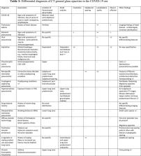
          Coronavirus disease 2019 (COVID-19), a recently emerged lower respiratory tract illness, has quickly become a pandemic. The purpose of this review is to discuss and differentiate COVID-19 typical imaging findings from other diseases, which can appear similar in the first instance. The typical CT findings of COVID-19 are bilateral and peripheral predominant ground glass opacities. As per the Fleischner Society consensus statement, CT is appropriate in certain scenarios, including patients who are at risk for and/or develop clinical worsening. The probability, that CT findings represent COVID-19, however, depends largely on the pre-test probability of infection, which is in turn defined by community prevalence of infection. When the community prevalence of COVID-19 is low, a large gap exists between positive predictive values of chest CT vs. RT-PCR. This implies that with usage of Chest CT there are a large number of false positive results. Imaging differentiation is important, for management and isolation purposes, and for appropriate disposition of CT false positive patients. We will discuss differential pathology with close imaging resemblance to typical CT imaging features of COVID-19, and, highlight CT features that may help in differentiation from other conditions.

          Related collections

          Most cited references71

          • Record: found
          • Abstract: found
          • Article: not found

          Clinical course and risk factors for mortality of adult inpatients with COVID-19 in Wuhan, China: a retrospective cohort study

          Summary Background Since December, 2019, Wuhan, China, has experienced an outbreak of coronavirus disease 2019 (COVID-19), caused by the severe acute respiratory syndrome coronavirus 2 (SARS-CoV-2). Epidemiological and clinical characteristics of patients with COVID-19 have been reported but risk factors for mortality and a detailed clinical course of illness, including viral shedding, have not been well described. Methods In this retrospective, multicentre cohort study, we included all adult inpatients (≥18 years old) with laboratory-confirmed COVID-19 from Jinyintan Hospital and Wuhan Pulmonary Hospital (Wuhan, China) who had been discharged or had died by Jan 31, 2020. Demographic, clinical, treatment, and laboratory data, including serial samples for viral RNA detection, were extracted from electronic medical records and compared between survivors and non-survivors. We used univariable and multivariable logistic regression methods to explore the risk factors associated with in-hospital death. Findings 191 patients (135 from Jinyintan Hospital and 56 from Wuhan Pulmonary Hospital) were included in this study, of whom 137 were discharged and 54 died in hospital. 91 (48%) patients had a comorbidity, with hypertension being the most common (58 [30%] patients), followed by diabetes (36 [19%] patients) and coronary heart disease (15 [8%] patients). Multivariable regression showed increasing odds of in-hospital death associated with older age (odds ratio 1·10, 95% CI 1·03–1·17, per year increase; p=0·0043), higher Sequential Organ Failure Assessment (SOFA) score (5·65, 2·61–12·23; p<0·0001), and d-dimer greater than 1 μg/mL (18·42, 2·64–128·55; p=0·0033) on admission. Median duration of viral shedding was 20·0 days (IQR 17·0–24·0) in survivors, but SARS-CoV-2 was detectable until death in non-survivors. The longest observed duration of viral shedding in survivors was 37 days. Interpretation The potential risk factors of older age, high SOFA score, and d-dimer greater than 1 μg/mL could help clinicians to identify patients with poor prognosis at an early stage. Prolonged viral shedding provides the rationale for a strategy of isolation of infected patients and optimal antiviral interventions in the future. Funding Chinese Academy of Medical Sciences Innovation Fund for Medical Sciences; National Science Grant for Distinguished Young Scholars; National Key Research and Development Program of China; The Beijing Science and Technology Project; and Major Projects of National Science and Technology on New Drug Creation and Development.
            Bookmark
            • Record: found
            • Abstract: found
            • Article: found

            Clinical Characteristics of 138 Hospitalized Patients With 2019 Novel Coronavirus–Infected Pneumonia in Wuhan, China

            In December 2019, novel coronavirus (2019-nCoV)-infected pneumonia (NCIP) occurred in Wuhan, China. The number of cases has increased rapidly but information on the clinical characteristics of affected patients is limited.
              Bookmark
              • Record: found
              • Abstract: found
              • Article: not found

              Time Course of Lung Changes On Chest CT During Recovery From 2019 Novel Coronavirus (COVID-19) Pneumonia

              Background Chest CT is used to assess the severity of lung involvement in COVID-19 pneumonia. Purpose To determine the change in chest CT findings associated with COVID-19 pneumonia from initial diagnosis until patient recovery. Materials and Methods This retrospective review included patients with RT-PCR confirmed COVID-19 infection presenting between 12 January 2020 to 6 February 2020. Patients with severe respiratory distress and/ or oxygen requirement at any time during the disease course were excluded. Repeat Chest CT was obtained at approximately 4 day intervals. The total CT score was the sum of lung involvement (5 lobes, score 1-5 for each lobe, range, 0 none, 25 maximum) was determined. Results Twenty one patients (6 males and 15 females, age 25-63 years) with confirmed COVID-19 pneumonia were evaluated. These patients under went a total of 82 pulmonary CT scans with a mean interval of 4±1 days (range: 1-8 days). All patients were discharged after a mean hospitalized period of 17±4 days (range: 11-26 days). Maximum lung involved peaked at approximately 10 days (with the calculated total CT score of 6) from the onset of initial symptoms (R2=0.25), p<0.001). Based on quartiles of patients from day 0 to day 26 involvement, 4 stages of lung CT were defined: Stage 1 (0-4 days): ground glass opacities (GGO) in 18/24 (75%) patients with the total CT score of 2±2; (2)Stage-2 (5-8d days): increased crazy-paving pattern 9/17 patients (53%) with a increase in total CT score (6±4, p=0.002); (3) Stage-3 (9-13days): consolidation 19/21 (91%) patients with the peak of total CT score (7±4); (4) Stage-4 (≥14 days): gradual resolution of consolidation 15/20 (75%) patients with a decreased total CT score (6±4) without crazy-paving pattern. Conclusion In patients recovering from COVID-19 pneumonia (without severe respiratory distress during the disease course), lung abnormalities on chest CT showed greatest severity approximately 10 days after initial onset of symptoms.
                Bookmark

                Author and article information

                Contributors
                Journal
                Radiology
                Radiology
                Radiology
                Radiology
                Radiological Society of North America
                0033-8419
                1527-1315
                07 July 2020
                : 202504
                Affiliations
                [1]From the Department of Radiology, Thomas Jefferson University Hospitals, 132 S 10th Street, 1079 Main Building, Philadelphia, Pa 19107 (M.P., A.D., R.B.); and Department of Radiology, University of Cincinnati Medical Center, 234 Goodman St, Cincinnati, OH (S.K.).
                Author notes
                Address correspondence to, A.D. (email: achala.donuru@ 123456jefferson.edu )

                Disclosures of Conflicts of Interest: M.P. disclosed no relevant relationships. A.D. disclosed no relevant relationships. R.B. disclosed no relevant relationships. S.K. disclosed no relevant relationships.

                Author information
                https://orcid.org/0000-0003-3308-918X
                https://orcid.org/0000-0001-8504-8764
                https://orcid.org/0000-0002-9819-7360
                Article
                202504
                10.1148/radiol.2020202504
                7350036
                32633678
                cc1cad59-1e0a-4dd6-b642-aa152a3b4832
                2020 by the Radiological Society of North America, Inc.

                This article is made available via the PMC Open Access Subset for unrestricted re-use and analyses in any form or by any means with acknowledgement of the original source. These permissions are granted for the duration of the COVID-19 pandemic or until permissions are revoked in writing. Upon expiration of these permissions, PMC is granted a perpetual license to make this article available via PMC and Europe PMC, consistent with existing copyright protections.

                History
                Categories
                Review
                Custom metadata
                yes

                Comments

                Comment on this article