4
views
0
recommends
+1 Recommend
0 collections
    0
    shares
      • Record: found
      • Abstract: found
      • Article: found
      Is Open Access

      Nursing Markedly Protects Postpartum Mice From Stroke: Associated Central and Peripheral Neuroimmune Changes and a Role for Oxytocin

      research-article

      Read this article at

      Bookmark
          There is no author summary for this article yet. Authors can add summaries to their articles on ScienceOpen to make them more accessible to a non-specialist audience.

          Abstract

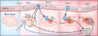
          Recent studies demonstrate significant neuroimmune changes in postpartum females, a period that also carries an increased risk of stroke. Oxytocin, a major hormone upregulated in the brains of nursing mothers, has been shown to both modulate neuroinflammation and protect against stroke. In the present study we assessed whether and how nursing modulates the neuroimmune response and injury after stroke. We observed that postpartum nursing mice were markedly protected from 1 h of transient middle cerebral artery occlusion (MCAO) relative to either non-pregnant/non-postpartum or non-nursing (pups removed) postpartum females. Nursing mice also expressed reduced levels of pro-inflammatory cytokines, had decreased migration of blood leukocytes into the brain following MCAO, and displayed peripheral neuroimmune changes characterized by increased spleen weight and increased fraction of spleen monocytes. Intranasal oxytocin treatment in non-pregnant females in part recapitulated the protective and anti-inflammatory effects associated with nursing. In summary, the results of the present study demonstrate that nursing in the postpartum period provides relative protection against transient ischemic stroke associated with decreased brain leukocytes and increased splenic monocytes. These effects appear to be regulated, at least in part, by oxytocin.

          Related collections

          Most cited references38

          • Record: found
          • Abstract: found
          • Article: found
          Is Open Access

          Inflammatory mechanisms in ischemic stroke: therapeutic approaches

          Acute ischemic stroke is the third leading cause of death in industrialized countries and the most frequent cause of permanent disability in adults worldwide. Despite advances in the understanding of the pathophysiology of cerebral ischemia, therapeutic options remain limited. Only recombinant tissue-plasminogen activator (rt-PA) for thrombolysis is currently approved for use in the treatment of this devastating disease. However, its use is limited by its short therapeutic window (three hours), complications derived essentially from the risk of hemorrhage, and the potential damage from reperfusion/ischemic injury. Two important pathophysiological mechanisms involved during ischemic stroke are oxidative stress and inflammation. Brain tissue is not well equipped with antioxidant defenses, so reactive oxygen species and other free radicals/oxidants, released by inflammatory cells, threaten tissue viability in the vicinity of the ischemic core. This review will discuss the molecular aspects of oxidative stress and inflammation in ischemic stroke and potential therapeutic strategies that target neuroinflammation and the innate immune system. Currently, little is known about endogenous counterregulatory immune mechanisms. However, recent studies showing that regulatory T cells are major cerebroprotective immunomodulators after stroke suggest that targeting the endogenous adaptive immune response may offer novel promising neuroprotectant therapies.
            Bookmark
            • Record: found
            • Abstract: found
            • Article: not found

            Inflammation and glial responses in ischemic brain lesions.

            Focal cerebral ischemia elicits a strong inflammatory response involving early recruitment of granulocytes and delayed infiltration of ischemic areas and the boundary zones by T cells and macrophages. Infiltration of hematogenous leukocytes is facilitated by an upregulation of the cellular adhesion molecules P-selectin, intercellular adhesion molecule-1 and vascular adhesion molecule-1 on endothelial cells. Blocking of the leukocyte/endothelial cell adhesion process significantly reduces stroke volume after transient, but not permanent middle cerebral artery occlusion. In the infarct region microglia are activated within hours and within days transform into phagocytes. Astrocytes upregulate intermediate filaments, synthesize neurotrophins and form glial scars. Local microglia and infiltrating macrophages demarcate infarcts and rapidly remove debris. Remote from the lesion no cellular infiltration occurs, but astroglia and microglia are transiently activated. Astrocytic activation is induced by spreading depression. In focal ischemia neurons die acutely by necrosis and in a delayed fashion by programmed cell death, apoptosis. Proinflammatory cytokines such as tumor necrosis factor-alpha and interleukin-1 beta are upregulated within hours in ischemic brain lesions. Either directly or via induction of neurotoxic mediators such as nitric oxide, cytokines may contribute to infarct progression in the post-ischemic period. On the other hand, inflammation is tightly linked with rapid removal of debris and repair processes. At present it is unclear whether detrimental effects of inflammation outweigh neuroprotective mechanisms or vice versa. In global ischemia inflammatory responses are limited, but micro- and astroglia are also strongly activated. Glial responses significantly differ between brain regions with selective neuronal death and neighbouring areas that are more resistent to ischemic damage.
              Bookmark
              • Record: found
              • Abstract: found
              • Article: not found

              Splenic atrophy in experimental stroke is accompanied by increased regulatory T cells and circulating macrophages.

              Induction of stroke not only produces local ischemia and brain damage, but also has profound effects on peripheral immune responses. In the current study, we evaluated effects on spleen and blood cells 4 days after stroke induction. Surprisingly, there was a less inflammatory cytokine profile in the middle cerebral artery occlusion-affected right brain hemisphere at 96 h compared with earlier time points. Moreover, our results demonstrate that stroke leads to splenic atrophy characterized by a reduction in organ size, a drastic loss of splenocyte numbers, and induction of annexin V+ and TUNEL+ cells within the spleen that are in the late stages of apoptosis. The consequence of this process was to reduce T cell proliferation responses and secretion of inflammatory cytokines, resulting in a state of profound immunosuppression. These changes produced a drastic reduction in B cell numbers in spleen and blood, and a novel increase in CD4+FoxP3+ regulatory T cells. Moreover, we detected a striking increase in the percentage of nonapoptotic CD11b+ VLA-4-negative macrophages/monocytes in blood. Immunosuppression in response to brain injury may account for the reduction of inflammatory factors in the stroke-affected brain, but also potentially could curtail protective immune responses in the periphery. These findings provide new evidence to support the contention that damage to the brain caused by cerebral ischemia provides a powerful negative signal to the peripheral immune system that ultimately induces a drastic state of immunosuppression caused by cell death as well as an increased presence of CD4+FoxP3+ regulatory T cells.
                Bookmark

                Author and article information

                Contributors
                Journal
                Front Neurosci
                Front Neurosci
                Front. Neurosci.
                Frontiers in Neuroscience
                Frontiers Media S.A.
                1662-4548
                1662-453X
                08 July 2019
                2019
                : 13
                : 609
                Affiliations
                [1] 1Department of Anesthesiology, Perioperative and Pain Medicine , Stanford, CA, United States
                [2] 2Department of Microbiology and Immunology , Stanford, CA, United States
                [3] 3Program in Immunology, Stanford University School of Medicine , Stanford, CA, United States
                Author notes

                Edited by: Ana Rosa Pérez, National Scientific and Technical Research Council (CONICET), Argentina

                Reviewed by: Benedetta Leuner, The Ohio State University, United States; Ana María Franchi, National Scientific and Technical Research Council (CONICET), Argentina

                *Correspondence: Creed M. Stary, cstary@ 123456stanford.edu

                This article was submitted to Neuroendocrine Science, a section of the journal Frontiers in Neuroscience

                Article
                10.3389/fnins.2019.00609
                6637858
                31354401
                ca1628e0-eb3a-42db-878a-693774fee0b5
                Copyright © 2019 Stary, Xu, Voloboueva, Alcántara-Hernández, Arvola, Idoyaga and Giffard.

                This is an open-access article distributed under the terms of the Creative Commons Attribution License (CC BY). The use, distribution or reproduction in other forums is permitted, provided the original author(s) and the copyright owner(s) are credited and that the original publication in this journal is cited, in accordance with accepted academic practice. No use, distribution or reproduction is permitted which does not comply with these terms.

                History
                : 27 March 2019
                : 28 May 2019
                Page count
                Figures: 6, Tables: 0, Equations: 0, References: 52, Pages: 10, Words: 0
                Funding
                Funded by: American Heart Association 10.13039/100000968
                Funded by: Suomen Kulttuurirahasto 10.13039/501100003125
                Funded by: National Institutes of Health 10.13039/100000002
                Categories
                Neuroscience
                Original Research

                Neurosciences
                mcao,facs,ischemia,innate,adaptive,focal ischemia,inflammation,cytokine
                Neurosciences
                mcao, facs, ischemia, innate, adaptive, focal ischemia, inflammation, cytokine

                Comments

                Comment on this article