46
views
0
recommends
+1 Recommend
0 collections
    0
    shares
      • Record: found
      • Abstract: found
      • Article: not found

      Baroreflex sensitivity differs among same strain Wistar rats from the same laboratory

      research-article

      Read this article at

      ScienceOpenPublisherPMC
      Bookmark
          There is no author summary for this article yet. Authors can add summaries to their articles on ScienceOpen to make them more accessible to a non-specialist audience.

          Abstract

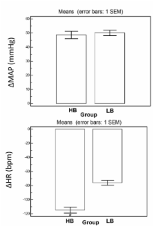
          Previous studies showed that a proportion of normotensive Sprague-Dawley rats spontaneously exhibit lower baroreflex sensitivity. However, investigations have not yet been carried out on Wistar rats. We aimed to compare baroreflex sensitivity among rats from the same strain and the same laboratory. Male Wistar normotensive rats (300–400g) were studied. Cannulas were inserted into the abdominal aortic artery through the right femoral artery to measure mean arterial pressure and heart rate. Baroreflex was calculated as the derivative of the variation of heart rate in function of the mean arterial pressure variation (ΔHR/ΔMAP) tested with a depressor dose of sodium nitroprusside (50 µg/kg) and with a pressor dose of phenylephrine (8µg/kg) in the right femoral venous approach through an inserted cannula. We divided the rats into four groups: i) high bradycardic baroreflex, baroreflex gain less than −2 tested with phenylephrine; ii) low bradycardic baroreflex, baroreflex gain between −1 and −2 tested with phenylephrine; iii) high tachycardic baroreflex, baroreflex gain less than −3 tested with sodium nitroprusside; and iv) low tachycardic baroreflex, baroreflex gain between −1 and −3 tested with sodium nitroprusside. Approximately 71% of the rats presented a decrease in bradycardic reflex while around half showed an increase in tachycardic reflex. No significant changes in basal mean arterial pressure and heart rate, tachycardic and bradycardic peak and heart rate range were observed. There was a significant change in baroreflex sensitivity among rats from the same strain and the same laboratory.

          Related collections

          Most cited references33

          • Record: found
          • Abstract: found
          • Article: not found

          Role of the medulla oblongata in hypertension.

          Brain pathways controlling arterial pressure are distributed throughout the neuraxis and are organized in topographically selective networks. In this brief review, we will focus on the medulla oblongata. The nucleus tractus solitarius (NTS) is the primary site of cardiorespiratory reflex integration. It is well accepted that lesions or other perturbations in the NTS can result in elevations of arterial pressure (AP), with many of the associated features so commonly found in humans. However, recent studies have shown 2 distinct subpopulations of neurons within the NTS that can influence AP in opposite ways. Commissural NTS neurons located on the midline may contribute to maintenance of hypertension in spontaneously hypertensive rats (SHR), because small lesions in this area result in a very significant reduction in AP. Also involved in this blood pressure regulation network are 2 distinct regions of the ventrolateral medulla: caudal (CVLM) and rostral (RVLM). Neurons in CVLM are thought to receive baroreceptor input and to relay rostrally to control the activity of the RVLM. Projections from CVLM to RVLM are inhibitory, and a lack of their activity may contribute to development of hypertension. The RVLM is critical to the tonic and reflexive regulation of AP. In different experimental models of hypertension, RVLM neurons receive significantly more excitatory inputs. This results in enhanced sympathetic neuronal activity, which is essential for the development and maintenance of the hypertension.
            Bookmark
            • Record: found
            • Abstract: found
            • Article: not found

            Arterial baroreflex modulation of heart rate in chronic heart failure: clinical and hemodynamic correlates and prognostic implications.

            In chronic heart failure (CHF), arterial baroreflex regulation of cardiac function is impaired, leading to a reduction in the tonic restraining influence on the sympathetic nervous system. Because baroreflex sensitivity (BRS), as assessed by the phenylephrine technique, significantly contributes to postinfarction risk stratification, the aim of the present study was to evaluate whether in CHF patients a depressed BRS is associated with a worse clinical hemodynamic status and unfavorable outcome. BRS was assessed in 282 CHF patients in sinus rhythm receiving stable medical therapy (age, 52+/-9 years; New York Heart Association [NYHA] class, 2.4+/-0.6; left ventricular ejection fraction [LVEF], 23+/-6%). The BRS of the entire population averaged 3.9+/-4.0 ms/mm Hg (mean+/-SD) and was significantly related to LVEF and hemodynamic parameters (LVEF, P 3 ms/mm Hg). During a mean follow-up of 15+/-12 months, 78 primary events (cardiac death, nonfatal cardiac arrest, and status 1 priority transplantation) occurred (27.6%). BRS was significantly related to outcome (log rank, 9.1; P<.01), with a relative risk of 2.7 (95% confidence interval, 1.6 to 4.7) for patients with the major derangement in BRS (<1.3 ms/mm Hg). At multivariate analysis, BRS was an independent predictor of death after adjustment for noninvasive known risk factors but not when hemodynamic indexes were also considered. In CHF patients with severe mitral regurgitation, however, BRS remained a strong prognostic marker independent of hemodynamic function. In moderate to severe CHF, a depressed sensitivity of vagal reflexes parallels the deterioration of clinical and hemodynamic status and is significantly associated with poor survival. Particularly in patients with severe mitral regurgitation the baroreceptor modulation of heart rate provides prognostic information of incremental value to hemodynamic parameters.
              Bookmark
              • Record: found
              • Abstract: found
              • Article: not found

              Brain renin-angiotensin system dysfunction in hypertension: recent advances and perspectives.

              This review focuses on the dysfunction of the intrinsic brain renin-angiotensin system (RAS) in the pathogenesis of hypertension. Hyperactivity of the brain RAS plays a critical role in mediating hypertension in both humans and animal models of hypertension, including the spontaneously hypertensive rat (SHR). The specific mechanisms by which increased brain RAS activity results in hypertension are not well understood but include increases in sympathetic vasomotor tone and impaired arterial baroreflex function. We discuss the contribution of endogenous angiotensin (Ang) II actions on presympathetic vasomotor rostral ventrolateral medulla neurons to enhance sympathetic activity and maintain hypertension. In addition, we discuss Ang II-induced attenuation of afferent baroreceptor feedback within the nucleus tractus solitarius and its relevance to the development of hypertension. We also outline the cellular and molecular mechanisms of Ang II signal transduction that may be critical for the initiation and establishment of hypertension. In particular, we present evidence for a phosphoinositide-3-kinase-dependent signaling pathway that appears to contribute to hypertension in the SHR, possibly via augmented Ang II-induced increases in neuronal firing rate and enhanced transcriptional noradrenaline neuromodulation. Finally, we outline future directions in utilizing our understanding of the brain RAS dysfunction in hypertension for the development of improved therapeutic intervention in hypertension.
                Bookmark

                Author and article information

                Journal
                Heart Int
                HI
                HI
                Heart International
                PAGEPress Publications (Pavia, Italy )
                1826-1868
                2036-2579
                29 September 2011
                29 September 2011
                : 6
                : 2
                : e9
                Affiliations
                [1 ]Departamento de Medicina, Disciplina de Cardiologia, Universidade Federal de São Paulo (UNIFESP), São Paulo, SP;
                [2 ]Laboratório de Escrita Científica, Departamento de Morfologia e Fisiologia, Faculdade de Medicina do ABC, Santo André, SP, Brazil
                Author notes
                Correspondence: Vitor E. Valenti, Departamento de Medicina, Disciplina de Cardiologia, Universidade Federal de São Paulo, UNIFESP, Rua Napoleão de Barros, 715 – Térreo, 04039-032, São Paulo, SP, Brazil. Tel. +55.11.5572-5462. E-mail: vitor.valenti@ 123456gmail.com

                Contributions: VEV, LCA, HMJ, ARPR, OFMF and CF participated in the acquisition of data and revision of the manuscript. VEV, LCA and CF conceived the study, determined the design, performed the statistical analysis, interpreted the data and drafted the manuscript. All authors read and gave final approval for the version submitted for publication.

                >Conflict of interest: the authors report no conflicts of interest.

                Article
                hi.2011.e9
                10.4081/hi.2011.e9
                3205781
                22049315
                c9e4f0b9-240b-4d04-9bb3-1b8a0c39c91c
                ©Copyright V.E. Valenti et al., 2011

                >This work is licensed under a Creative Commons Attribution NonCommercial 3.0 License (CC BY-NC 3.0).

                Licensee PAGEPress, Italy

                History
                : 09 May 2011
                : 29 August 2011
                : 09 September 2011
                Categories
                Laboratory Investigation

                Cardiovascular Medicine
                sympathetic nerve.,parasympathetic nerve,autonomic nervous system,hypertension,baroreceptors

                Comments

                Comment on this article