1
views
0
recommends
+1 Recommend
0 collections
    0
    shares
      • Record: found
      • Abstract: found
      • Article: found
      Is Open Access

      First in patient assessment of brain tumor infiltrative margins using simultaneous time-resolved measurements of 5-ALA-induced PpIX fluorescence and tissue autofluorescence

      research-article

      Read this article at

      Bookmark
          There is no author summary for this article yet. Authors can add summaries to their articles on ScienceOpen to make them more accessible to a non-specialist audience.

          Abstract.

          Significance: 5-aminolevulinic acid (5-ALA)-induced protoporphyrin IX (PpIX) fluorescence is currently used for image-guided glioma resection. Typically, this widefield imaging method highlights the bulk of high-grade gliomas, but it underperforms at the infiltrating edge where PpIX fluorescence is not visible to the eyes. Fluorescence lifetime imaging (FLIm) has the potential to detect PpIX fluorescence below the visible detection threshold. Moreover, simultaneous acquisition of time-resolved nicotinamide adenine (phosphate) dinucleotide [NAD(P)H] fluorescence may provide metabolic information from the tumor environment to further improve overall tumor detection.

          Aim: We investigate the ability of pulse sampling, fiber-based FLIm to simultaneously image PpIX and NAD(P)H fluorescence of glioma infiltrative margins in patients.

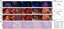
          Approach: A mesoscopic fiber-based point-scanning FLIm device (355 nm pulses) was used to simultaneously resolve the fluorescence decay of PpIX (629/53 nm) and NAD(P)H (470/28 nm). The FLIm device enabled data acquisition at room light and rapid ( < 33    ms ) augmentation of FLIm parameters on the surgical field-of-view. FLIm measurements from superficial tumors and tissue areas around the resection margins were performed on three glioblastoma patients in vivo following inspection of PpIX visible fluorescence with a conventional neurosurgical microscope. Microbiopsies were collected from FLIm imaged areas for histopathological evaluation.

          Results: The average lifetime from PpIX and NAD(P)H fluorescence distinguished between tumor and surrounding tissue. FLIm measurements of resection margins presented a range of PpIX and NAD(P)H lifetime values ( τ PpIX 3 to 14 ns, τ NAD ( P ) H = 3 to 6 ns) associated with unaffected tissue and areas of low-density tumor infiltration.

          Conclusions: Intraoperative FLIm could simultaneously detect the emission of PpIX and NAD(P)H from patients in vivo during craniotomy procedures. This approach doubles as a clinical tool to identify tumor areas while performing tissue resection and as a research tool to study tumor microenvironmental changes in vivo. Intraoperative FLIm of 5-ALA-induced PpIX and tissue autofluorescence makes a promising surgical adjunct to guide tumor resection surgery.

          Related collections

          Most cited references26

          • Record: found
          • Abstract: found
          • Article: not found

          The phasor approach to fluorescence lifetime imaging analysis.

          Changing the data representation from the classical time delay histogram to the phasor representation provides a global view of the fluorescence decay at each pixel of an image. In the phasor representation we can easily recognize the presence of different molecular species in a pixel or the occurrence of fluorescence resonance energy transfer. The analysis of the fluorescence lifetime imaging microscopy (FLIM) data in the phasor space is done observing clustering of pixels values in specific regions of the phasor plot rather than by fitting the fluorescence decay using exponentials. The analysis is instantaneous since is not based on calculations or nonlinear fitting. The phasor approach has the potential to simplify the way data are analyzed in FLIM, paving the way for the analysis of large data sets and, in general, making the FLIM technique accessible to the nonexpert in spectroscopy and data analysis.
            Bookmark
            • Record: found
            • Abstract: found
            • Article: found
            Is Open Access

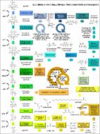
            Fatty acid oxidation is required for the respiration and proliferation of malignant glioma cells

            Background. Glioma is the most common form of primary malignant brain tumor in adults, with approximately 4 cases per 100 000 people each year. Gliomas, like many tumors, are thought to primarily metabolize glucose for energy production; however, the reliance upon glycolysis has recently been called into question. In this study, we aimed to identify the metabolic fuel requirements of human glioma cells. Methods. We used database searches and tissue culture resources to evaluate genotype and protein expression, tracked oxygen consumption rates to study metabolic responses to various substrates, performed histochemical techniques and fluorescence-activated cell sorting-based mitotic profiling to study cellular proliferation rates, and employed an animal model of malignant glioma to evaluate a new therapeutic intervention. Results. We observed the presence of enzymes required for fatty acid oxidation within human glioma tissues. In addition, we demonstrated that this metabolic pathway is a major contributor to aerobic respiration in primary-cultured cells isolated from human glioma and grown under serum-free conditions. Moreover, inhibiting fatty acid oxidation reduces proliferative activity in these primary-cultured cells and prolongs survival in a syngeneic mouse model of malignant glioma. Conclusions. Fatty acid oxidation enzymes are present and active within glioma tissues. Targeting this metabolic pathway reduces energy production and cellular proliferation in glioma cells. The drug etomoxir may provide therapeutic benefit to patients with malignant glioma. In addition, the expression of fatty acid oxidation enzymes may provide prognostic indicators for clinical practice.
              Bookmark
              • Record: found
              • Abstract: found
              • Article: found
              Is Open Access

              Metabolic Reprogramming in Glioma

              Many cancers have long been thought to primarily metabolize glucose for energy production—a phenomenon known as the Warburg Effect, after the classic studies of Otto Warburg in the early twentieth century. Yet cancer cells also utilize other substrates, such as amino acids and fatty acids, to produce raw materials for cellular maintenance and energetic currency to accomplish cellular tasks. The contribution of these substrates is increasingly appreciated in the context of glioma, the most common form of malignant brain tumor. Multiple catabolic pathways are used for energy production within glioma cells, and are linked in many ways to anabolic pathways supporting cellular function. For example: glycolysis both supports energy production and provides carbon skeletons for the synthesis of nucleic acids; meanwhile fatty acids are used both as energetic substrates and as raw materials for lipid membranes. Furthermore, bio-energetic pathways are connected to pro-oncogenic signaling within glioma cells. For example: AMPK signaling links catabolism with cell cycle progression; mTOR signaling contributes to metabolic flexibility and cancer cell survival; the electron transport chain produces ATP and reactive oxygen species (ROS) which act as signaling molecules; Hypoxia Inducible Factors (HIFs) mediate interactions with cells and vasculature within the tumor environment. Mutations in the tumor suppressor p53, and the tricarboxylic acid cycle enzymes Isocitrate Dehydrogenase 1 and 2 have been implicated in oncogenic signaling as well as establishing metabolic phenotypes in genetically-defined subsets of malignant glioma. These pathways critically contribute to tumor biology. The aim of this review is two-fold. Firstly, we present the current state of knowledge regarding the metabolic strategies employed by malignant glioma cells, including aerobic glycolysis; the pentose phosphate pathway; one-carbon metabolism; the tricarboxylic acid cycle, which is central to amino acid metabolism; oxidative phosphorylation; and fatty acid metabolism, which significantly contributes to energy production in glioma cells. Secondly, we highlight processes (including the Randle Effect, AMPK signaling, mTOR activation, etc.) which are understood to link bio-energetic pathways with oncogenic signals, thereby allowing the glioma cell to achieve a pro-malignant state.
                Bookmark

                Author and article information

                Contributors
                Journal
                J Biomed Opt
                J Biomed Opt
                JBOPFO
                JBO
                Journal of Biomedical Optics
                Society of Photo-Optical Instrumentation Engineers
                1083-3668
                1560-2281
                2 February 2022
                February 2022
                2 February 2022
                : 27
                : 2
                : 020501
                Affiliations
                [a ]University of California Davis , Department of Biomedical Engineering, Davis, California, United States
                [b ]University of California Davis , Department of Pathology and Laboratory Medicine, Sacramento, California, United States
                [c ]University of California Davis , Department of Neurological Surgery, Sacramento, California, United States
                Author notes
                [* ]Address all correspondence to Laura Marcu, lmarcu@ 123456ucdavis.edu
                Author information
                https://orcid.org/0000-0002-6748-3751
                https://orcid.org/0000-0002-6715-5176
                https://orcid.org/0000-0003-1222-4071
                https://orcid.org/0000-0002-5003-6322
                https://orcid.org/0000-0001-6790-0888
                Article
                JBO-210294LRR 210294LRR
                10.1117/1.JBO.27.2.020501
                8809358
                35112514
                c938c8ff-2516-4513-9026-3241a3bbd9bb
                © 2022 The Authors

                Published by SPIE under a Creative Commons Attribution 4.0 International License. Distribution or reproduction of this work in whole or in part requires full attribution of the original publication, including its DOI.

                History
                : 16 September 2021
                : 11 January 2022
                Page count
                Figures: 3, Tables: 0, References: 26, Pages: 8
                Funding
                Funded by: National Institutes of Health
                Award ID: R01CA250512
                Award ID: R21CA252510
                Categories
                JBO Letters
                Letter
                Custom metadata
                Alfonso-García et al.: First in patient assessment of brain tumor infiltrative margins using simultaneous…

                Biomedical engineering
                fluorescence lifetime imaging,5-ala-induced ppix fluorescence,nicotinamide adenine (phosphate) dinucleotide,brain tumor,glioblastoma

                Comments

                Comment on this article