There is no author summary for this article yet. Authors can add summaries to their articles on ScienceOpen to make them more accessible to a non-specialist audience.
Abstract
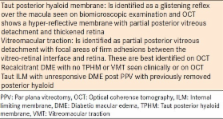
Surgery for late complications of proliferative diabetic retinopathy remains the cornerstone
of management even in patients who have received optimal laser photocoagulation and
medical therapy. With improvisation in the surgical techniques and development of
micro-incision surgical techniques for vitrectomy, the indications for surgical intervention
are expanding to include diabetic macular edema with a greater number of patients
undergoing early intervention. This review describes the current indications, surgical
techniques, adjunctive anti-vascular endothelial growth factor therapy, surgical outcomes,
and postoperative complications of pars plana vitrectomy for proliferative diabetic
retinopathy and macular edema.
Pars plana vitrectomy with separation of the posterior hyaloid was performed in 10 eyes with diabetic macular edema and traction associated with a thickened and taut premacular posterior hyaloid. Nine of the 10 eyes had previous macular photocoagulation. Preoperative fluorescein angiography showed a deep and diffuse pattern of leakage in the macula. Intraoperatively, the attached and thickened posterior hyaloid was lifted and separated from the retina. Postoperatively, vision improved in nine eyes. The macular traction and edema resolved in eight eyes and decreased in two. Complications included a vitreous hemorrhage, a rhegmatogenous retinal detachment, cataract formation, and a mild epimacular membrane, each occurring in one eye. Vitreous surgery can improve the visual prognosis of some eyes with diabetic macular traction and edema associated with a thickened and taut posterior hyaloid.
To evaluate vitrectomy for diabetic macular edema (DME) in eyes with at least moderate vision loss and vitreomacular traction. Prospective cohort study. The primary cohort included 87 eyes with DME and vitreomacular traction based on investigator's evaluation, visual acuity 20/63-20/400, optical coherence tomography (OCT) central subfield >300 microns and no concomitant cataract extraction at the time of vitrectomy. Surgery was performed according to the investigator's usual routine. Follow-up visits were performed after 3 months, 6 months (primary end point), and 1 year. Visual acuity, OCT retinal thickening, and operative complications. At baseline, median visual acuity in the 87 eyes was 20/100 and median OCT thickness was 491 microns. During vitrectomy, additional procedures included epiretinal membrane peeling in 61%, internal limiting membrane peeling in 54%, panretinal photocoagulation in 40%, and injection of corticosteroids at the close of the procedure in 64%. At 6 months, median OCT central subfield thickness decreased by 160 microns, with 43% having central subfield thickness or =10 letters in 38% (95% confidence interval, 28%-49%) and deteriorated by > or =10 letters in 22% (95% confidence interval, 13%-31%). Postoperative complications through 6 months included vitreous hemorrhage (5 eyes), elevated intraocular pressure requiring treatment (7 eyes), retinal detachment (3 eyes), and endophthalmitis (1 eye). Few changes in results were noted between 6 months and 1 year. After vitrectomy performed for DME and vitreomacular traction, retinal thickening was reduced in most eyes. Between 28% and 49% of eyes with characteristics similar to those included in this study are likely to have improvement of visual acuity, whereas between 13% and 31% are likely to have worsening. The operative complication rate is low and similar to what has been reported for this procedure. These data provide estimates of surgical outcomes and serve as a reference for future studies that might consider vitrectomy for DME in eyes with at least moderate vision loss and vitreomacular traction. Copyright 2010 American Academy of Ophthalmology. Published by Elsevier Inc. All rights reserved.
Analyses of visual acuity and visual field results in the Diabetic Retinopathy Study provide evidence that photocoagulation treatment as carried out according to the study protocol (extensive "scatter" photocoagulation and focal treatment of new vessels) is of benefit in preventing severe visual loss, over a two-year follow-up period, in eyes with proliferative retinopathy. Location of new vessels relative to the disk, severity on new vessels, and the presence of hemorrhage (vitreous or preretinal) all proved to be important prognostic factors. On the basis of these findings, these steps have been taken: All patients in the study have been informed of results to date and given an explanation of their implications. Photocoagulation treatment will be considered for the initially untreated eyes which now or in the future fulfill any one of the following criteria: (a) moderate or severe new vessels on or within 1-disk diameter of the optic disk; (b) mild new vessels on or within 1-disk diameter of the optic disk if fresh hemorrhage is present; and (c) moderate or severe new vessels elsewhere, if fresh hemorrhage is present. Follow-up of all patients will continue to allow long-term comparison between the argon- and xenon-treatment techniques employed. Further analyses of accumulating data will be performed to evaluate more completely the efficacy of photocoagulation therapy.
Journal ID (iso-abbrev): Middle East Afr J Ophthalmol
Journal ID (publisher-id): MEAJO
Title:
Middle East African Journal of Ophthalmology
Publisher:
Medknow Publications & Media Pvt Ltd
(India
)
ISSN
(Print):
0974-9233
ISSN
(Electronic):
0975-1599
Publication date
(Print):
Oct-Dec 2013
Volume: 20
Issue: 4
Pages: 283-292
Affiliations
[1
]Vitreoretinal and Uveitis Division, King Khaled Eye Specialist Hospital, Riyadh, Kingdom
of Saudi Arabia
[2
]Retina Division, Wilmer Eye Institute, Johns Hopkins University School of Medicine,
Baltimore, MD, USA
Author notes
Corresponding Author: Dr. Vishali Gupta, The King Khaled Eye Specialist Hospital, Al-Oruba Street, PO Box
7191, Riyadh 11462, Kingdom of Saudi Arabia,. E-mail:
vishalisara@
123456yahoo.co.in
This is an open-access article distributed under the terms of the Creative Commons
Attribution-Noncommercial-Share Alike 3.0 Unported, which permits unrestricted use,
distribution, and reproduction in any medium, provided the original work is properly
cited.
scite shows how a scientific paper has been cited by providing the context of the citation, a classification describing whether it supports, mentions, or contrasts the cited claim, and a label indicating in which section the citation was made.