19
views
0
recommends
+1 Recommend
0 collections
    0
    shares
      • Record: found
      • Abstract: found
      • Article: found
      Is Open Access

      Machine learning-based analysis of a semi-automated PI-RADS v2.1 scoring for prostate cancer

      research-article

      Read this article at

      Bookmark
          There is no author summary for this article yet. Authors can add summaries to their articles on ScienceOpen to make them more accessible to a non-specialist audience.

          Abstract

          Background

          Prostate Imaging-Reporting and Data System version 2.1 (PI-RADS v2.1) was developed to standardize the interpretation of multiparametric MRI (mpMRI) for prostate cancer (PCa) detection. However, a significant inter-reader variability among radiologists has been found in the PI-RADS assessment. The purpose of this study was to evaluate the diagnostic performance of an in-house developed semi-automated model for PI-RADS v2.1 scoring using machine learning methods.

          Methods

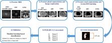
          The study cohort included an MRI dataset of 59 patients (PI-RADS v2.1 score 2 = 18, score 3 = 10, score 4 = 16, and score 5 = 15). The proposed semi-automated model involved prostate gland and zonal segmentation, 3D co-registration, lesion region of interest marking, and lesion measurement. PI-RADS v2.1 scores were assessed based on lesion measurements and compared with the radiologist PI-RADS assessment. Machine learning methods were used to evaluate the diagnostic accuracy of the proposed model by classification of PI-RADS v2.1 scores.

          Results

          The semi-automated PI-RADS assessment based on the proposed model correctly classified 50 out of 59 patients and showed a significant correlation ( r = 0.94, p < 0.05) with the radiologist assessment. The proposed model achieved an accuracy of 88.00% ± 0.98% and an area under the receiver-operating characteristic curve (AUC) of 0.94 for score 2 vs. score 3 vs. score 4 vs. score 5 classification and accuracy of 93.20 ± 2.10% and AUC of 0.99 for low score vs. high score classification using fivefold cross-validation.

          Conclusion

          The proposed semi-automated PI-RADS v2.1 assessment system could minimize the inter-reader variability among radiologists and improve the objectivity of scoring.

          Related collections

          Most cited references27

          • Record: found
          • Abstract: found
          • Article: not found

          Global cancer statistics 2020: GLOBOCAN estimates of incidence and mortality worldwide for 36 cancers in 185 countries

          This article provides an update on the global cancer burden using the GLOBOCAN 2020 estimates of cancer incidence and mortality produced by the International Agency for Research on Cancer. Worldwide, an estimated 19.3 million new cancer cases (18.1 million excluding nonmelanoma skin cancer) and almost 10.0 million cancer deaths (9.9 million excluding nonmelanoma skin cancer) occurred in 2020. Female breast cancer has surpassed lung cancer as the most commonly diagnosed cancer, with an estimated 2.3 million new cases (11.7%), followed by lung (11.4%), colorectal (10.0 %), prostate (7.3%), and stomach (5.6%) cancers. Lung cancer remained the leading cause of cancer death, with an estimated 1.8 million deaths (18%), followed by colorectal (9.4%), liver (8.3%), stomach (7.7%), and female breast (6.9%) cancers. Overall incidence was from 2-fold to 3-fold higher in transitioned versus transitioning countries for both sexes, whereas mortality varied <2-fold for men and little for women. Death rates for female breast and cervical cancers, however, were considerably higher in transitioning versus transitioned countries (15.0 vs 12.8 per 100,000 and 12.4 vs 5.2 per 100,000, respectively). The global cancer burden is expected to be 28.4 million cases in 2040, a 47% rise from 2020, with a larger increase in transitioning (64% to 95%) versus transitioned (32% to 56%) countries due to demographic changes, although this may be further exacerbated by increasing risk factors associated with globalization and a growing economy. Efforts to build a sustainable infrastructure for the dissemination of cancer prevention measures and provision of cancer care in transitioning countries is critical for global cancer control.
            Bookmark
            • Record: found
            • Abstract: found
            • Article: not found

            Prostate Imaging Reporting and Data System Version 2.1: 2019 Update of Prostate Imaging Reporting and Data System Version 2

            The Prostate Imaging Reporting and Data System version 2 (PI-RADS v2) was developed with a consensus-based process using a combination of published data, and expert observations and opinions. In the short time since its release, numerous studies have validated the value of PI-RADS v2 but, as expected, have also identified a number of ambiguities and limitations, some of which have been documented in the literature with potential solutions offered. To address these issues, the PI-RADS Steering Committee, again using a consensus-based process, has recommended several modifications to PI-RADS v2, maintaining the framework of assigning scores to individual sequences and using these scores to derive an overall assessment category. This updated version, described in this article, is termed PI-RADS v2.1. It is anticipated that the adoption of these PI-RADS v2.1 modifications will improve inter-reader variability and simplify PI-RADS assessment of prostate magnetic resonance imaging even further. Research on the value and limitations on all components of PI-RADS v2.1 is strongly encouraged.
              Bookmark
              • Record: found
              • Abstract: found
              • Article: not found

              Active contours without edges.

              We propose a new model for active contours to detect objects in a given image, based on techniques of curve evolution, Mumford-Shah (1989) functional for segmentation and level sets. Our model can detect objects whose boundaries are not necessarily defined by the gradient. We minimize an energy which can be seen as a particular case of the minimal partition problem. In the level set formulation, the problem becomes a "mean-curvature flow"-like evolving the active contour, which will stop on the desired boundary. However, the stopping term does not depend on the gradient of the image, as in the classical active contour models, but is instead related to a particular segmentation of the image. We give a numerical algorithm using finite differences. Finally, we present various experimental results and in particular some examples for which the classical snakes methods based on the gradient are not applicable. Also, the initial curve can be anywhere in the image, and interior contours are automatically detected.
                Bookmark

                Author and article information

                Contributors
                Journal
                Front Oncol
                Front Oncol
                Front. Oncol.
                Frontiers in Oncology
                Frontiers Media S.A.
                2234-943X
                24 November 2022
                2022
                : 12
                : 961985
                Affiliations
                [1] 1 Centre for Biomedical Engineering, Indian Institute of Technology Delhi , New Delhi, India
                [2] 2 Department of Nuclear Magnetic Resonance (NMR), All India Institute of Medical Sciences , New Delhi, India
                [3] 3 Department of Radiodiagnosis, All India Institute of Medical Sciences , New Delhi, India
                [4] 4 Department of Biomedical Engineering, All India Institute of Medical Sciences , New Delhi, India
                Author notes

                Edited by: Antonella Santone, University of Molise, Italy

                Reviewed by: Francesco Mercaldo, University of Molise, Italy; Caterina Gaudiano, Sant’Orsola-Malpighi Polyclinic, Italy

                *Correspondence: Amit Mehndiratta, amit.mehndiratta@ 123456keble.oxon.org

                This article was submitted to Cancer Imaging and Image-directed Interventions, a section of the journal Frontiers in Oncology

                Article
                10.3389/fonc.2022.961985
                9730331
                36505875
                c8a68c36-f4a6-4152-ab8e-17ce4b96ac2b
                Copyright © 2022 Singh, Kumar, Das, Singh and Mehndiratta

                This is an open-access article distributed under the terms of the Creative Commons Attribution License (CC BY). The use, distribution or reproduction in other forums is permitted, provided the original author(s) and the copyright owner(s) are credited and that the original publication in this journal is cited, in accordance with accepted academic practice. No use, distribution or reproduction is permitted which does not comply with these terms.

                History
                : 05 June 2022
                : 27 October 2022
                Page count
                Figures: 4, Tables: 3, Equations: 6, References: 28, Pages: 10, Words: 4528
                Categories
                Oncology
                Original Research

                Oncology & Radiotherapy
                lesion measurement,prostate cancer,prostate imaging-reporting and data system version 2.1,machine learning,mri

                Comments

                Comment on this article