99
views
0
recommends
+1 Recommend
0 collections
    0
    shares
      • Record: found
      • Abstract: found
      • Article: found
      Is Open Access

      Parapapillary Atrophy: Histological Gamma Zone and Delta Zone

      research-article

      Read this article at

      Bookmark
          There is no author summary for this article yet. Authors can add summaries to their articles on ScienceOpen to make them more accessible to a non-specialist audience.

          Abstract

          Background

          To examine histomorphometrically the parapapillary region in human eyes.

          Methodology/Principal Findings

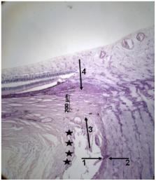
          The histomorphometric study included 65 human globes (axial length:21–37 mm). On anterior-posterior histological sections, we measured the distance Bruch's membrane end (BME)-optic nerve margin (“Gamma zone”), BME-retinal pigment epithelium (RPE) (“Beta zone”), BME-beginning of non-occluded choriocapillaris, and BME-beginning of photoreceptor layer. “Delta zone” was defined as part of gamma zone in which blood vessels of at least 50 µm diameter were not present over a length of >300 µm. Beta zone (mean length:0.35±0.52 mm) was significantly (P = 0.01) larger in the glaucoma group than in the non-glaucomatous group. It was not significantly (P = 0.28) associated with axial length. Beta zone was significantly (P = 0.004) larger than the region with occluded choriocapillaris. Gamma zone (mean length:0.63±1.25 mm) was associated with axial length ( P<0.001;r 2 = 0.73) with an increase starting at an axial length of 26.5 mm. It was not significantly ( P = 0.24) associated with glaucomatous optic neuropathy. Delta zone (present only in eyes with axial length of ≥27 mm) was associated with axial length ( P = 0.001) and scleral flange length ( P<0.001) but not with glaucoma ( P = 0.73).

          Conclusions/Significance

          Parapapillary gamma zone (peripapillary sclera without overlying choroid, Bruch's membrane and deep retinal layers) was related with axial globe elongation and was independent of glaucoma. Delta zone (no blood vessels >50 µm diameter within gamma zone) was present only in highly axially elongated globes and was not related with glaucoma. Beta zone (Bruch's membrane without RPE) was correlated with glaucoma but not with globe elongation. Since the region with occluded choriocapillaris was smaller than beta zone, complete loss of RPE may have occurred before complete choriocapillaris closure.

          Related collections

          Most cited references14

          • Record: found
          • Abstract: found
          • Article: not found

          Factors influencing optic nerve head biomechanics.

          The biomechanical environment within the optic nerve head (ONH) may play a role in retinal ganglion cell loss in glaucomatous optic neuropathy. This was a systematic analysis in which finite element methods were used to determine which anatomic and biomechanical factors most influenced the biomechanical response of the ONH to acute changes in IOP. Based on a previously described computational model of the eye, each of 21 input factors, representing the biomechanical properties of relevant ocular tissues, the IOP, and 14 geometric factors were independently varied. The biomechanical response of the ONH tissues was quantified through a set of 29 outcome measures, including peak and mean stress and strain within each tissue, and measures of geometric changes in ONH tissues. Input factors were ranked according to their aggregated influence on groups of outcome measures. The five input factors that had the largest influence across all outcome measures were, in ranked order: stiffness of the sclera, radius of the eye, stiffness of the lamina cribrosa, IOP, and thickness of the scleral shell. The five least influential factors were, in reverse ranked order: retinal thickness, peripapillary rim height, cup depth, cup-to-disc ratio, and pial thickness. Factor ranks were similar for various outcome measure groups and factor ranges. The model predicts that ONH biomechanics are strongly dependent on scleral biomechanical properties. Acute deformations of ONH tissues, and the consequent high levels of neural tissue strain, were less strongly dependent on the action of IOP directly on the internal surface of the ONH than on the indirect effects of IOP on the sclera. This suggests that interindividual variations in scleral properties could be a risk factor for the development of glaucoma. Eye size and lamina cribrosa biomechanical properties also have a strong influence on ONH biomechanics.
            Bookmark
            • Record: found
            • Abstract: found
            • Article: not found

            Enhanced depth imaging optical coherence tomography of deep optic nerve complex structures in glaucoma.

            To assess the usefulness of enhanced depth imaging (EDI) optical coherence tomography (OCT) for evaluating deep structures of the optic nerve complex (ONC; optic nerve head and peripapillary structures) in glaucoma. Prospective, observational study. Seventy-three established glaucoma patients (139 eyes) with a range of glaucomatous damage. Serial horizontal and vertical EDI OCT images of the ONC were obtained from both eyes of each participant. Deep ONC structures, including the lamina cribrosa (LC), short posterior ciliary artery (SPCA), central retinal artery (CRA), central retinal vein (CRV), peripapillary choroid and sclera, and subarachnoid space around the optic nerve, were investigated for their visibility and morphologic features. Deep ONC structures identified in EDI OCT images. Visual field mean deviation of 139 included eyes was -11.8 ± 8.6 dB (range, -28.70 to -2.01 dB). The anterior laminar surface was identified in all eyes in the central laminar area and in 91 (65%) eyes in the periphery beneath the neuroretinal and scleral rims or vascular structures. The LC pores with various shapes and sizes were visualized in 106 (76%) eyes, mainly in the central and temporal areas of the LC. Localized LC lesions seen on optic disc photographs were identified as focal LC defects (partial loss of LC tissue) in the EDI OCT images. The locations of the CRA and CRV were identified in all eyes. In the LC, the CRA maintained a straight shape with a consistent caliber, but the CRV (and tributaries) assumed a more irregular shape. The SPCAs, their branches through the emissary canals in the sclera, or both were visualized in 120 (86%) eyes. The subarachnoid space around the optic nerve was identified with varying degrees of clarity in 25 eyes (18%): 17 had high myopia and extensive parapapillary atrophy. Intrachoroidal cavitation or choroidal schisis, which had been unrecognized clinically, was identified in 2 eyes (1%) with high myopia. Enhanced depth imaging OCT was able to visualize a wide variety of deep ONC structures in glaucoma patients and may be helpful in detecting, conceptualizing, and understanding basic and complicated in vivo anatomic and pathologic features of the ONC in glaucoma. Proprietary or commercial disclosure may be found after the references. Copyright © 2012 American Academy of Ophthalmology. Published by Elsevier Inc. All rights reserved.
              Bookmark
              • Record: found
              • Abstract: found
              • Article: not found

              Parapapillary chorioretinal atrophy in normal and glaucoma eyes. I. Morphometric data.

              Glaucomatous optic nerve damage is associated with alterations of the intra- and parapapillary optic disc area. We measured and compared the parapapillary region in 582 eyes of 321 patients suffering from chronic primary open-angle glaucoma and in 390 eyes of 231 normal subjects. Only one randomly assessed eye per patient and subject was taken for statistical analysis. Highly mopic eyes with a myopic refractive error of more than -8.00 diopters had been excluded. The parapapillary chorioretinal atrophy was divided into a peripheral zone "Alpha" with irregular hyper- and hypopigmentation, and a more central zone "Beta" characterized by whitish colour, visible large choroidal vessels and visible sclera. In the normal eyes both zones were significantly (P less than 0.001) largest and most common in the temporal horizontal sector, followed by the inferior temporal sector, the superior temporal sector and finally the nasal sector. In the glaucoma group both zones were significantly larger (P less than 0.0001; Mann-Whitney test) and more frequent than in the normal eyes (0.40 +/- 0.32 mm2 versus 0.65 +/- 0.49 mm2 for zone Alpha, 0.13 +/- 0.42 mm2 versus 0.79 +/- 1.17 mm2 for zone Beta). The differences were significant also for the earliest glaucoma stage of this study. They were most marked for the nasal parapapillary sector. Significant differences (P less than 0.001) between the normal group and the earliest glaucoma stage were: zone Alpha larger than 0.20 mm2 or broader than 0.20 mm in the temporal horizontal sector, total area of zone Alpha larger than 0.30 mm2, occurrence of zone Alpha in the nasal sector, and occurrence of zone Beta anywhere.
                Bookmark

                Author and article information

                Contributors
                Role: Editor
                Journal
                PLoS One
                PLoS ONE
                plos
                plosone
                PLoS ONE
                Public Library of Science (San Francisco, USA )
                1932-6203
                2012
                18 October 2012
                : 7
                : 10
                : e47237
                Affiliations
                [1 ]Department of Ophthalmology, Medical Faculty Mannheim of the Ruprecht-Karls-University Heidelberg, Germany
                [2 ]Department of Ophthalmology, Friedrich-Alexander University Erlangen-Nürnberg, Erlangen, Germany
                [3 ]Department of Ophthalmology and Vision Science, Eye and ENT Hospital, Shanghai Medical College, Fudan University, Shanghai, China
                [4 ]State Key Laboratory of Medical Neurobiology, Institutes of Brain Science, Fudan University, Shanghai, China
                University of Sydney, Australia
                Author notes

                Competing Interests: The authors have declared that no competing interests exist.

                Conceived and designed the experiments: JBJ SBJ RAJ LH YD XS SP-J. Performed the experiments: JBJ SBJ RAJ SP-J. Analyzed the data: JBJ SBJ RAJ LH YD XS SP-J. Contributed reagents/materials/analysis tools: JBJ LH. Wrote the paper: JBJ SBJ RAJ LH YD XS SP-J.

                Article
                PONE-D-12-20006
                10.1371/journal.pone.0047237
                3475708
                23094040
                c7efcd3a-a1d6-434c-964c-e4650435b970
                Copyright @ 2012

                This is an open-access article distributed under the terms of the Creative Commons Attribution License, which permits unrestricted use, distribution, and reproduction in any medium, provided the original author and source are credited.

                History
                : 10 July 2012
                : 10 September 2012
                Page count
                Pages: 7
                Funding
                No current external funding sources for this study.
                Categories
                Research Article
                Biology
                Anatomy and Physiology
                Neurological System
                Neuroanatomy
                Ocular System
                Ocular Anatomy
                Medicine
                Anatomy and Physiology
                Neurological System
                Neuroanatomy
                Ocular System
                Ocular Anatomy
                Ophthalmology
                Glaucoma
                Surgery
                Ophthalmology

                Uncategorized
                Uncategorized

                Comments

                Comment on this article