14
views
0
recommends
+1 Recommend
0 collections
    0
    shares
      • Record: found
      • Abstract: found
      • Article: found
      Is Open Access

      Overview of RAW264.7 for osteoclastogensis study: Phenotype and stimuli

      review-article

      Read this article at

      Bookmark
          There is no author summary for this article yet. Authors can add summaries to their articles on ScienceOpen to make them more accessible to a non-specialist audience.

          Abstract

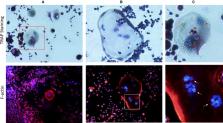
          Bone homeostasis is preserved by the balance of maintaining between the activity of osteogenesis and osteoclastogenesis. However, investigations for the osteoclastogenesis were hampered by considerable difficulties associated with isolating and culturing osteoclast in vivo. As the alternative, stimuli‐induced osteoclasts formation from RAW264.7 cells (RAW‐OCs) have gain its importance for extensively osteoclastogenic study of bone diseases, such as rheumatoid arthritis, osteoporosis, osteolysis and periodontitis. However, considering the RAW‐OCs have not yet been well‐characterized and RAW264.7 cells are polymorphic because of a diverse phenotype of the individual cells comprising this cell linage, and different fate associated with various stimuli contributions. Thus, in present study, we provide an overview for current knowledge of the phenotype of RAW264.7 cells, as well as the current understanding of the complicated interactions between various stimuli and RAW‐OCs in the light of the recent progress.

          Related collections

          Most cited references125

          • Record: found
          • Abstract: found
          • Article: found
          Is Open Access

          Bone marrow CD169+ macrophages promote the retention of hematopoietic stem and progenitor cells in the mesenchymal stem cell niche

          Hematopoietic stem cells (HSCs) reside in specialized bone marrow (BM) niches regulated by the sympathetic nervous system (SNS). Here, we have examined whether mononuclear phagocytes modulate the HSC niche. We defined three populations of BM mononuclear phagocytes that include Gr-1hi monocytes (MOs), Gr-1lo MOs, and macrophages (MΦ) based on differential expression of Gr-1, CD115, F4/80, and CD169. Using MO and MΦ conditional depletion models, we found that reductions in BM mononuclear phagocytes led to reduced BM CXCL12 levels, the selective down-regulation of HSC retention genes in Nestin+ niche cells, and egress of HSCs/progenitors to the bloodstream. Furthermore, specific depletion of CD169+ MΦ, which spares BM MOs, was sufficient to induce HSC/progenitor egress. MΦ depletion also enhanced mobilization induced by a CXCR4 antagonist or granulocyte colony-stimulating factor. These results highlight two antagonistic, tightly balanced pathways that regulate maintenance of HSCs/progenitors in the niche during homeostasis, in which MΦ cross talk with the Nestin+ niche cell promotes retention, and in contrast, SNS signals enhance egress. Thus, strategies that target BM MΦ hold the potential to augment stem cell yields in patients that mobilize HSCs/progenitors poorly.
            Bookmark
            • Record: found
            • Abstract: found
            • Article: not found

            Tumor necrosis factor receptor family member RANK mediates osteoclast differentiation and activation induced by osteoprotegerin ligand.

            A receptor that mediates osteoprotegerin ligand (OPGL)-induced osteoclast differentiation and activation has been identified via genomic analysis of a primary osteoclast precursor cell cDNA library and is identical to the tumor necrosis factor receptor (TNFR) family member RANK. The RANK mRNA was highly expressed by isolated bone marrow-derived osteoclast progenitors and by mature osteoclasts in vivo. Recombinant OPGL binds specifically to RANK expressed by transfected cell lines and purified osteoclast progenitors. Transgenic mice expressing a soluble RANK-Fc fusion protein have severe osteopetrosis because of a reduction in osteoclasts, similar to OPG transgenic mice. Recombinant RANK-Fc binds with high affinity to OPGL in vitro and blocks osteoclast differentiation and activation in vitro and in vivo. Furthermore, polyclonal Ab against the RANK extracellular domain promotes osteoclastogenesis in bone marrow cultures suggesting that RANK activation mediates the effects of OPGL on the osteoclast pathway. These data indicate that OPGL-induced osteoclastogenesis is directly mediated through RANK on osteoclast precursor cells.
              Bookmark
              • Record: found
              • Abstract: found
              • Article: found
              Is Open Access

              Macrophage Polarisation: an Immunohistochemical Approach for Identifying M1 and M2 Macrophages

              Macrophage polarization is increasingly recognised as an important pathogenetic factor in inflammatory and neoplastic diseases. Proinflammatory M1 macrophages promote T helper (Th) 1 responses and show tumoricidal activity. M2 macrophages contribute to tissue repair and promote Th2 responses. CD68 and CD163 are used to identify macrophages in tissue sections. However, characterisation of polarised macrophages in situ has remained difficult. Macrophage polarisation is regulated by transcription factors, pSTAT1 and RBP-J for M1, and CMAF for M2. We reasoned that double-labelling immunohistochemistry for the detection of macrophage markers together with transcription factors may be suitable to characterise macrophage polarisation in situ. To test this hypothesis, we have studied conditions associated with Th1- and Th2-predominant immune responses: infectious mononucleosis and Crohn’s disease for Th1 and allergic nasal polyps, oxyuriasis, wound healing and foreign body granulomas for predominant Th2 response. In all situations, CD163+ cells usually outnumbered CD68+ cells. Moreover, CD163+ cells, usually considered as M2 macrophages, co-expressing pSTAT1 and RBP-J were found in all conditions examined. The numbers of putative M1 macrophages were higher in Th1- than in Th2-associated diseases, while more M2 macrophages were seen in Th2- than in Th1 related disorders. In most Th1-related diseases, the balance of M1 over M2 cells was shifted towards M1 cells, while the reverse was observed for Th2-related conditions. Hierarchical cluster analysis revealed two distinct clusters: cluster I included Th1 diseases together with cases with high numbers of CD163+pSTAT1+, CD68+pSTAT1+, CD163+RBP-J+ and CD68+RBP-J+ macrophages; cluster II comprised Th2 conditions together with cases displaying high numbers of CD163+CMAF+ and CD68+CMAF+ macrophages. These results suggest that the detection of pSTAT1, RBP-J, and CMAF in the context of CD68 or CD163 expression is a suitable tool for the characterisation of macrophage polarisation in situ. Furthermore, CD163 cannot be considered a reliable M2 marker when used on its own.
                Bookmark

                Author and article information

                Contributors
                lingbokong@163.com
                Journal
                J Cell Mol Med
                J. Cell. Mol. Med
                10.1111/(ISSN)1582-4934
                JCMM
                Journal of Cellular and Molecular Medicine
                John Wiley and Sons Inc. (Hoboken )
                1582-1838
                1582-4934
                20 March 2019
                May 2019
                : 23
                : 5 ( doiID: 10.1111/jcmm.2019.23.issue-5 )
                : 3077-3087
                Affiliations
                [ 1 ] Department of Spine, School of Medicine Honghui‐hospital, Xi’an Jiaotong University Xi’an China
                [ 2 ] Department of Neuroscience Johns Hopkins University Baltimore Maryland
                Author notes
                [*] [* ] Correspondence

                Lingbo Kong, Department of Spine, School of Medicine, Honghui‐Hospital, Xi’an Jiaotong University, Xi’an, China.

                Email: lingbokong@ 123456163.com

                Author information
                https://orcid.org/0000-0002-3655-3171
                Article
                JCMM14277
                10.1111/jcmm.14277
                6484317
                30892789
                c7d6a9fa-c3d8-40a6-8e42-02c26392e8e2
                © 2019 The Authors. Journal of Cellular and Molecular Medicine published by John Wiley & Sons Ltd and Foundation for Cellular and Molecular Medicine.

                This is an open access article under the terms of the http://creativecommons.org/licenses/by/4.0/ License, which permits use, distribution and reproduction in any medium, provided the original work is properly cited.

                History
                : 24 January 2019
                : 13 February 2019
                : 16 February 2019
                Page count
                Figures: 3, Tables: 0, Pages: 11, Words: 8667
                Funding
                Funded by: China Postdoctoral Science Foundation
                Award ID: 2018T111458
                Funded by: Shannxi Postdoctoral Science Foundation
                Categories
                Review
                Reviews
                Custom metadata
                2.0
                jcmm14277
                May 2019
                Converter:WILEY_ML3GV2_TO_NLMPMC version:5.6.2.1 mode:remove_FC converted:26.04.2019

                Molecular medicine
                bone marrow macrophage,lps,osteoclast,phenotype,rankl,raw264.7,tnf‐a
                Molecular medicine
                bone marrow macrophage, lps, osteoclast, phenotype, rankl, raw264.7, tnf‐a

                Comments

                Comment on this article