16
views
0
recommends
+1 Recommend
0 collections
    0
    shares
      • Record: found
      • Abstract: found
      • Article: found
      Is Open Access

      Simultaneous Confocal Scanning Laser Ophthalmoscopy Combined with High-Resolution Spectral-Domain Optical Coherence Tomography: A Review

      review-article

      Read this article at

      Bookmark
          There is no author summary for this article yet. Authors can add summaries to their articles on ScienceOpen to make them more accessible to a non-specialist audience.

          Abstract

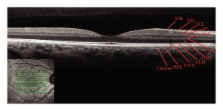
          We aimed to evaluate technical aspects and the clinical relevance of a simultaneous confocal scanning laser ophthalmoscope and a high-speed, high-resolution, spectral-domain optical coherence tomography (SDOCT) device for retinal imaging. The principle of confocal scanning laser imaging provides a high resolution of retinal and choroidal vasculature with low light exposure. Enhanced contrast, details, and image sharpness are generated using confocality. The real-time SDOCT provides a new level of accuracy for assessment of the angiographic and morphological correlation. The combined system allows for simultaneous recordings of topographic and tomographic images with accurate correlation between them. Also it can provide simultaneous multimodal imaging of retinal pathologies, such as fluorescein and indocyanine green angiographies, infrared and blue reflectance (red-free) images, fundus autofluorescence images, and OCT scans (Spectralis HRA + OCT; Heidelberg Engineering, Heidelberg, Germany). The combination of various macular diagnostic tools can lead to a better understanding and improved knowledge of macular diseases.

          Related collections

          Most cited references55

          • Record: found
          • Abstract: found
          • Article: not found

          Optical coherence tomography.

          A technique called optical coherence tomography (OCT) has been developed for noninvasive cross-sectional imaging in biological systems. OCT uses low-coherence interferometry to produce a two-dimensional image of optical scattering from internal tissue microstructures in a way that is analogous to ultrasonic pulse-echo imaging. OCT has longitudinal and lateral spatial resolutions of a few micrometers and can detect reflected signals as small as approximately 10(-10) of the incident optical power. Tomographic imaging is demonstrated in vitro in the peripapillary area of the retina and in the coronary artery, two clinically relevant examples that are representative of transparent and turbid media, respectively.
            Bookmark
            • Record: found
            • Abstract: found
            • Article: not found

            A pilot study of enhanced depth imaging optical coherence tomography of the choroid in normal eyes.

            To measure macular choroidal thickness in normal eyes at different points using enhanced depth imaging (EDI) optical coherence tomography (OCT) and to evaluate the association of choroidal thickness and age. Retrospective, observational case series. EDI OCT images were obtained in patients without significant retinal or choroidal pathologic features. The images were obtained by positioning a spectral-domain OCT device close enough to the eye to acquire an inverted image. Seven sections were obtained within a 5 x 30-degree area centered at the fovea, with 100 scans averaged for each section. The choroid was measured from the outer border of the retinal pigment epithelium to the inner scleral border at 500-microm intervals of a horizontal section from 3 mm temporal to the fovea to 3 mm nasal to the fovea. Statistical analysis was performed to evaluate variations of choroidal thickness at each location and to correlate choroidal thickness and patient age. The mean age of the 30 patients (54 eyes) was 50.4 years (range, 19 to 85 years), and 14 patients (46.7%) were female. The choroid was thickest underneath the fovea (mean, 287 microm; standard deviation, +/- 76 microm). Choroidal thickness decreased rapidly in the nasal direction and averaged 145 microm (+/- 57 microm) at 3 mm nasal to the fovea. Increasing age was correlated significantly with decreasing choroidal thickness at all points measured. Regression analysis suggested that the subfoveal choroidal thickness decreased by 15.6 microm for each decade of life. Choroidal thickness seems to vary topographically within the posterior pole. The thickness of the choroid showed a negative correlation with age. The decrease in the thickness of the choroid may play a role in the pathophysiologic features of various age-related ocular conditions.
              Bookmark
              • Record: found
              • Abstract: found
              • Article: not found

              Optical coherence tomography of the human retina.

              To demonstrate optical coherence tomography for high-resolution, noninvasive imaging of the human retina. Optical coherence tomography is a new imaging technique analogous to ultrasound B scan that can provide cross-sectional images of the retina with micrometer-scale resolution. Survey optical coherence tomographic examination of the retina, including the macula and optic nerve head in normal human subjects. Research laboratory. Convenience sample of normal human subjects. Correlation of optical coherence retinal tomographs with known normal retinal anatomy. Optical coherence tomographs can discriminate the cross-sectional morphologic features of the fovea and optic disc, the layered structure of the retina, and normal anatomic variations in retinal and retinal nerve fiber layer thicknesses with 10-microns depth resolution. Optical coherence tomography is a potentially useful technique for high depth resolution, cross-sectional examination of the fundus.
                Bookmark

                Author and article information

                Journal
                J Ophthalmol
                JOP
                Journal of Ophthalmology
                Hindawi Publishing Corporation
                2090-004X
                2090-0058
                2011
                1 November 2011
                : 2011
                : 743670
                Affiliations
                1Retina Service, Department of Ophthalmology, Federal University of São Paulo, 04021-001 São Paulo, SP, Brazil
                2Department of Ophthalmology, University of Bonn, 53012 Bonn, Germany
                Author notes
                *Verônica Castro Lima: vecastrolima@ 123456yahoo.com.br

                Academic Editor: Fernando Penha

                Article
                10.1155/2011/743670
                3206361
                22132313
                c790e2df-8829-47cd-8395-2aacac4710cf
                Copyright © 2011 Verônica Castro Lima et al.

                This is an open access article distributed under the Creative Commons Attribution License, which permits unrestricted use, distribution, and reproduction in any medium, provided the original work is properly cited.

                History
                : 23 June 2011
                : 17 August 2011
                Categories
                Review Article

                Ophthalmology & Optometry
                Ophthalmology & Optometry

                Comments

                Comment on this article