- Record: found
- Abstract: not found
- Article: not found
A case report of vaccine-induced immune thrombocytopenia and thrombosis syndrome after Ad26.COV2.S vaccine (Janssen/Johnson & Johnson) ☆
letter
Maxime Castan
a
,
* ,
Marlène Damin-Pernik
b ,
Guillaume Thiéry
c
,
d
,
e ,
Dominique Page
c ,
David M. Smadja
f
,
g ,
Laurent Bertoletti
h
,
i
,
j
31 January 2022
Read this article at

There is no author summary for this article yet. Authors can add summaries to their articles on ScienceOpen to make them more accessible to a non-specialist audience.
Abstract
Abbreviations
COVID
coronavirus disease
DIC
disseminated intravascular coagulation
FDPs
fibrin degradation products
ITP
immune thrombocytopenic purpura
MRI
magnetic resonance imaging
PCR
polymerase chain reaction
PF4
platelet factor 4
SARS-CoV-2
severe acute respiratory syndrome coronavirus 2
TTP
thrombotic thrombocytopenic purpura
VITT
vaccine-induced immune thrombocytopenia and thrombosis
Introduction
Vaccine-induced immune thrombocytopenia and thrombosis (VITT) syndrome has recently
been described after the ChAdOx1 nCoV-19 vaccine (AstraZeneca) [1]. This syndrome
is characterized by the occurrence of venous and/or arterial thrombosis, often at
atypical sites, with thrombocytopenia and positive anti-PF4 (platelet factor 4) antibodies,
in a recent context of vaccination against coronavirus disease 2019 (COVID-19).
We describe here a case of VITT syndrome, which occurred following vaccination with
Ad26.COV2.S vaccine (Janssen).
Case report
On August 2, 2021, ten days after receiving a dose of Ad26.COV2.S vaccine (Janssen/Johnson
& Johnson), a 57-years-old man was admitted for left hemiplegia. The rest of clinical
examination was unremarkable. Severe acute respiratory syndrome coronavirus 2 (SARS-CoV-2)
polymerase chain reaction (PCR) testing by nasopharyngeal swab was negative. He has
no significant medical history and does not take any long-term treatment. Ischemic
stroke, of thromboembolic origin with description of a proximal occlusion of the right
internal carotid artery, was confirmed on brain magnetic resonance imaging (MRI).
Initial blood tests were abnormal, including thrombocytopenia at 27 G/L, hepatic cytolysis
at 10N and biological disseminated intravascular coagulation (DIC) with fibrinogen < 1 g/L,
D-dimer> 128,000 ng/mL and fibrin degradation products (FDPs) > 150 μg/mL. Myelogram
was normal.
Arterial Doppler ultrasound of the supra-aortic trunks confirmed a complete thrombosis
of the right internal carotid artery. Ultrasound and abdomino-pelvic CT scan revealed
partial portal vein thrombosis and right and middle hepatic vein thrombosis. Pain
in the left leg prompted the realization of a venous Doppler ultrasound of the lower
limbs, finding a distal deep venous thrombosis. Transthoracic echocardiography was
normal.
Patient received intravenous acetylsalicylic acid (250 mg/24 h) and subcutaneous enoxaparin
(100 IU/kg/12 h) and was admitted to the intensive care unit.
Neurological examination showed cognitive disorders, hemiparesis of the left upper
limb rated at 1/5 and hemiparesis of the left lower limb side at 2/5, with signs of
spatial neglect. Because of neurological worsening (appearance of a left homonymous
hemianopsia at 48 hours), brain CT scan showed intracranial bleeding leading to stop
antithrombotic agent and curative anticoagulation.
VITT syndrome was suspected. Differential diagnostics were ruled out (SARS-CoV-2 infection,
others infections, immune thrombocytopenic purpura (ITP), drugs, hypersplenism, genetic
disorder, cancer, trauma, surgery, immobilization, thrombotic thrombocytopenic purpura
(TTP), thrombophilia). Search for anti-PF4 antibodies and a platelet aggregation test
were performed, from which only anti-PF4 antibodies returned positive at 1,181 IU/L
(N < 0.5) by ELISA method (Zymutest HIA IgGAM Hyphen), platelet aggregation test returned
normal.
The patient received corticosteroids 0,75 mg/kg and intravenous immunoglobulins at
2 g/kg over 2 days, either seven days after the onset of symptoms. Biological parameters
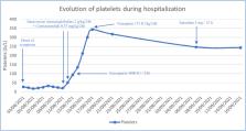
improved over the next few days, in particular platelets (Fig. 1
) and fibrinogen which returned to normal values in 5 days and liver function tests
in 17 days. On day 10, internal carotid artery was re-permeabilized on arterial Doppler
ultrasound, and thrombus completely disappeared on the control a month and a half
later.
Figure 1
Evolution of platelets during hospitalization.
Concomitantly, neurological symptoms began to improve, including hemiplegia, cognitive
and ophthalmologic disorders. Follow-up brain scan did not show any new intracranial
bleeding. Preventive anticoagulation by subcutaneous enoxaparin 4000 IU/24 h was reinitiated,
followed by subcutaneous tinzaparin 175 IU/kg/24 h and later by Apixaban 5 mg/12 h,
once the liver function is normal.
Seven days after initiation of treatment, neurological examination improved, with
hemiparesis of the left upper limb rated at 3/5 and hemiparesis of the left lower
limb rated at 4/5.
Two months after the onset of symptoms, neurological examination objectified hemiparesis
of the left upper limb rated at 4/5 and hemiparesis of the left lower limb rated at
4/5.
Four months after the onset of symptoms, patient can walk a short distance with a
cane.
Discussion
According to us, this is the first case of VITT syndrome reported to the French Regional
Pharmacovigilance Centers in France for the Ad26.COV2.S vaccine (Janssen/Johnson &
Johnson). A declaration to the French National Pharmacovigilance Database was made
on August 9, 2021 and was registered under number SE20212123. Causality relationship
between Ad26.COV2.S vaccine (Janssen/Johnson & Johnson) and VITT syndrome was assessed
as “likely” (I3, C2S3) with the French pharmacovigilance causality [2]. The latest
report of pharmacovigilance of ChAdOx1 nCoV-19 (AstraZeneca) on November 25, 2021 described
29 cases of confirmed VITT in France vs 4 cases for the Ad26.COV2.S vaccine (Janssen/Johnson
& Johnson) [3].
The diagnosis of VITT is definite according to the consensus of the UK Haematology
Expert Group [4] with a delay of onset of symptoms of 10 days after vaccination, multiple
thrombosis even if the sites described are not the most frequent, biological assessment
with a major DIC (D-dimer > 4000 ng/mL, platelets at 27 G/L) having been resolved
few days after initiation of immunoglobulins and corticosteroids and positive anti-PF4 antibodies
ELISA assay.
Our research in the literature found several studies concerning mainly ChAdOx1 nCoV-19
(AstraZeneca) on this syndrome in the United States and in Europe in particular in
the United Kingdom, in Denmark, in Norway, in Austria and in Germany. Locations described
as being the most frequent were cerebral veins, pulmonary arteries and multiple sites
[4]. Although similar, there are differences between VITT syndrome induced by ChAdOx1 nCoV-19
(AstraZeneca) and Ad26.COV2.S vaccine (Janssen/Johnson & Johnson): in particular median
time to onset of, respectively, 10- and 16-days post-vaccination and lower D-dimer
levels in Ad26.COV2.S vaccine recipients [5]. There would also be more intracerebral
hemorrhages after Ad26.COV2.S administration (Janssen/Johnson & Johnson) [5]. These
differences are important to consider in the diagnostic process of VITT syndrome.
Incidence was around 1/50,000-100,000 for both vaccines [4], [6] but there is a higher
incidence of ChAdOx1 nCoV-19 (AstraZeneca) in the United Kingdom, a country where
this vaccine was mainly used, unlike in the United States where Ad26.COV2.S vaccine
(Janssen/Johnson & Johnson) is the majority. The fact that the incidence of occurrence
of VITT syndrome is lower in recipients of Ad26.COV2.S vaccine (Janssen/Johnson &
Johnson) may be explained by the later release and by less use than other vaccines.
Treatments were variable and mainly included corticosteroids and intravenous immunoglobulins.
Other treatments have been tested, specifically rituximab (anti-CD20) and eculizumab
(anti factor C5) [7], the principle remaining of slowing down immune response [7].
It was not recommended to have recourse to platelet transfusions except to cover any
possible procedures, as this would promote aggravation of thrombosis [4], [8].
Mortality reported in the literature varied from 23% to 72% depending on the existence
or not of intracranial bleeding and thrombocytopenia < 30 G/L [4], [5], and also associated
with early diagnosis and rapid initiation of appropriate treatment. A predictive mortality
score has been developed: the FAPIC score [9]. It includes fibrinogen (< 1,5 g/L),
age (≤ 60 years), platelet count (< 25 G/L), intracerebral hemorrhage and cerebral
venous thrombosis, and can be used to predict mortality of VITT syndrome [9].
In our patient's case, platelets normalized quickly after initiation of treatment.
Due to the description of a non-heparin-dependent pathophysiological mechanism [8],
we anticoagulated the patient with heparin treatment, and this did not cause a significant
drop in platelets, which remained at a normal level.
Conclusion
As of 10 November 2021, there have been more than 7 billion doses of vaccine worldwide
and currently available vaccines have been extensively tested in clinical trials and
their efficacy and safety is well established. Common vaccine-related side effects
are fever, myalgia, arthralgia and headache [8]. Occurrence of serious adverse events
attributable to the vaccine therefore remains difficult to interpret. VITT syndrome
has only been reported very few times in the literature [1], [4], [6], [8], [9], [10],
around 474 cases for the ChAdOx1 nCov-19 in European Union and United Kingdom on October
9, 2021, and 28 cases for the Ad26.COV2.S vaccine in USA on July 19, 2021. Risk-benefit
ratio remains in favor of vaccination, in particular since SARS-CoV-2 infection is
more thrombogenic than vaccination [6].
Link between occurrence of VITT syndrome and adenovirus-vector-based SARS-CoV-2 vaccines
is increasingly established, but this event remains rare and it therefore appears
essential to identify the VITT syndrome early on: implementation of rapid treatment
allows almost immediate clinical improvement and would therefore reduce mortality
of this extremely serious adverse event.
Disclosure of interest
The authors declare that they have no competing interest.
Related collections
Most cited references9
- Record: found
- Abstract: found
- Article: not found
Thrombotic Thrombocytopenia after ChAdOx1 nCov-19 Vaccination
- Record: found
- Abstract: found
- Article: found
Pathologic Antibodies to Platelet Factor 4 after ChAdOx1 nCoV-19 Vaccination
Marie Scully, Deepak Singh, Robert Lown … (2021)
- Record: found
- Abstract: found
- Article: not found
Clinical Features of Vaccine-Induced Immune Thrombocytopenia and Thrombosis
Sue Pavord, Marie Scully, Beverley J. Hunt … (2021)