7
views
0
recommends
+1 Recommend
0 collections
    0
    shares
      • Record: found
      • Abstract: found
      • Article: found
      Is Open Access

      TLR4 promotes microglial pyroptosis via lncRNA-F630028O10Rik by activating PI3K/AKT pathway after spinal cord injury

      research-article

      Read this article at

      Bookmark
          There is no author summary for this article yet. Authors can add summaries to their articles on ScienceOpen to make them more accessible to a non-specialist audience.

          Abstract

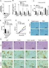
          Neuroinflammation plays a crucial role in the secondary phase of spinal cord injury (SCI), and is initiated following the activation of toll-like receptor 4 (TLR4). However, the downstream mechanism remains unknown. Pyroptosis is a form of inflammatory programmed cell death, which is closely involved in neuroinflammation, and it can be regulated by TLR4 according to a recent research. In addition, several studies have shown that long non-coding RNAs (lncRNAs) based mechanisms were related to signal transduction downstream of TLR4 in the regulation of inflammation. Thus, in this study, we want to determine whether TLR4 can regulate pyroptosis after SCI via lncRNAs. Our results showed that TLR4 was activated following SCI and promoted the expression of lncRNA-F630028O10Rik. This lncRNA functioned as a ceRNA for miR-1231-5p/Col1a1 axis and enhanced microglial pyroptosis after SCI by activating the PI3K/AKT pathway. Furthermore, we determined STAT1 was the upstream transcriptional factor of IncRNA-F630028O10Rik and was induced by the damage-responsive TLR4/MyD88 signal. Our findings provide new insights and a novel therapeutic strategy for treating SCI.

          Related collections

          Most cited references42

          • Record: found
          • Abstract: found
          • Article: not found

          Pyroptosis.

          Injury and physical trauma may inflict accidental cell death, but we have come to realize during the past four decades that cells may also actively engage cell death when needed. These regulated cell death forms are intrinsically connected with human embryonic development, homeostatic maintenance and disease pathology. For instance, the human body is composed of approximately 10(14) cells, millions of which are removed daily by apoptosis and replaced with newly differentiated cells in order to secure organ functionality. Apoptotic cells are orderly packed in 'apoptotic bodies' for uptake by neighboring cells and professional phagocytes, thereby avoiding deleterious inflammatory responses by circulating leukocytes. Unlike apoptosis, however, more recently identified forms of regulated cell death - such as necroptosis and pyroptosis - are characterized by an early breach of the plasma membrane integrity, which results in extracellular spilling of the intracellular contents. Here, we will describe and discuss this and other features of pyroptosis.
            Bookmark
            • Record: found
            • Abstract: found
            • Article: not found

            Inflammasomes in the CNS.

            Microglia and macrophages in the CNS contain multimolecular complexes termed inflammasomes. Inflammasomes function as intracellular sensors for infectious agents as well as for host-derived danger signals that are associated with neurological diseases, including meningitis, stroke and Alzheimer's disease. Assembly of an inflammasome activates caspase 1 and, subsequently, the proteolysis and release of the cytokines interleukin-1β and interleukin-18, as well as pyroptotic cell death. Since the discovery of inflammasomes in 2002, there has been burgeoning recognition of their complexities and functions. Here, we review the current understanding of the functions of different inflammasomes in the CNS and their roles in neurological diseases.
              Bookmark
              • Record: found
              • Abstract: found
              • Article: not found

              Inhibiting NF-κB activation by small molecules as a therapeutic strategy.

              Because nuclear factor-κB (NF-κB) is a ubiquitously expressed proinflammatory transcription factor that regulates the expression of over 500 genes involved in cellular transformation, survival, proliferation, invasion, angiogenesis, metastasis, and inflammation, the NF-κB signaling pathway has become a potential target for pharmacological intervention. A wide variety of agents can activate NF-κB through canonical and noncanonical pathways. Canonical pathway involves various steps including the phosphorylation, ubiquitination, and degradation of the inhibitor of NF-κB (IκBα), which leads to the nuclear translocation of the p50-p65 subunits of NF-κB followed by p65 phosphorylation, acetylation and methylation, DNA binding, and gene transcription. Thus, agents that can inhibit protein kinases, protein phosphatases, proteasomes, ubiquitination, acetylation, methylation, and DNA binding steps have been identified as NF-κB inhibitors. Because of the critical role of NF-κB in cancer and various chronic diseases, numerous inhibitors of NF-κB have been identified. In this review, however, we describe only small molecules that suppress NF-κB activation, and the mechanism by which they block this pathway. Copyright © 2010 Elsevier B.V. All rights reserved.
                Bookmark

                Author and article information

                Contributors
                mxs_huashan@163.com
                lfzsubmission@163.com
                Journal
                Cell Death Dis
                Cell Death Dis
                Cell Death & Disease
                Nature Publishing Group UK (London )
                2041-4889
                10 August 2020
                10 August 2020
                August 2020
                : 11
                : 8
                : 693
                Affiliations
                [1 ]GRID grid.8547.e, ISNI 0000 0001 0125 2443, Department of Orthopedics, , Shanghai Fifth People’s Hospital, Fudan University, ; Shanghai, 200240 China
                [2 ]GRID grid.411405.5, ISNI 0000 0004 1757 8861, Department of Orthopedics, , Huashan Hospital, Fudan University, ; Shanghai, 200040 China
                Author information
                http://orcid.org/0000-0001-8887-5554
                Article
                2824
                10.1038/s41419-020-02824-z
                7443136
                32826878
                c6dd367d-f9e2-4882-be7c-a79f54a4ee98
                © The Author(s) 2020

                Open Access This article is licensed under a Creative Commons Attribution 4.0 International License, which permits use, sharing, adaptation, distribution and reproduction in any medium or format, as long as you give appropriate credit to the original author(s) and the source, provide a link to the Creative Commons license, and indicate if changes were made. The images or other third party material in this article are included in the article’s Creative Commons license, unless indicated otherwise in a credit line to the material. If material is not included in the article’s Creative Commons license and your intended use is not permitted by statutory regulation or exceeds the permitted use, you will need to obtain permission directly from the copyright holder. To view a copy of this license, visit http://creativecommons.org/licenses/by/4.0/.

                History
                : 21 February 2020
                : 21 July 2020
                : 23 July 2020
                Funding
                Funded by: FundRef https://doi.org/10.13039/501100001809, National Natural Science Foundation of China (National Science Foundation of China);
                Award ID: 81772385
                Award ID: 81871522
                Award ID: 81871522
                Award Recipient :
                Funded by: Shanghai Sailing Program, 20YF1438100
                Funded by: Shanghai Sailing Program, 20YF1429900
                Categories
                Article
                Custom metadata
                © The Author(s) 2020

                Cell biology
                epigenetics,neuroimmunology
                Cell biology
                epigenetics, neuroimmunology

                Comments

                Comment on this article