10
views
0
recommends
+1 Recommend
0 collections
    0
    shares
      • Record: found
      • Abstract: found
      • Article: found
      Is Open Access

      Dysregulated IER3 Expression is Associated with Enhanced Apoptosis in Titin-Based Dilated Cardiomyopathy

      research-article

      Read this article at

      Bookmark
          There is no author summary for this article yet. Authors can add summaries to their articles on ScienceOpen to make them more accessible to a non-specialist audience.

          Abstract

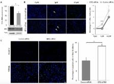
          Apoptosis (type I programmed cell death) of cardiomyocytes is a major process that plays a role in the progression of heart failure. The early response gene IER3 regulates apoptosis in a wide variety of cells and organs. However, its role in heart failure is largely unknown. Here, we investigate the role of IER3 in an inducible heart failure mouse model. Heart failure was induced in a mouse model that imitates a human titin truncation mutation we found in a patient with dilated cardiomyopathy (DCM). Transferase dUTP nick end labeling (TUNEL) and ssDNA stainings showed induction of apoptosis in titin-deficient cardiomyocytes during heart failure development, while IER3 response was dysregulated. Chromatin immunoprecipitation and knock-down experiments revealed that IER3 proteins target the promotors of anti-apoptotic genes and act as an anti-apoptotic factor in cardiomyocytes. Its expression is blunted during heart failure development in a titin-deficient mouse model. Targeting the IER3 pathway to reduce cardiac apoptosis might be an effective therapeutic strategy to combat heart failure.

          Related collections

          Most cited references31

          • Record: found
          • Abstract: found
          • Article: not found

          HL-1 cells: a cardiac muscle cell line that contracts and retains phenotypic characteristics of the adult cardiomyocyte.

          We have derived a cardiac muscle cell line, designated HL-1, from the AT-1 mouse atrial cardiomyocyte tumor lineage. HL-1 cells can be serially passaged, yet they maintain the ability to contract and retain differentiated cardiac morphological, biochemical, and electrophysiological properties. Ultrastructural characteristics typical of embryonic atrial cardiac muscle cells were found consistently in the cultured HL-1 cells. Reverse transcriptase-PCR-based analyses confirmed a pattern of gene expression similar to that of adult atrial myocytes, including expression of alpha-cardiac myosin heavy chain, alpha-cardiac actin, and connexin43. They also express the gene for atrial natriuretic factor. Immunohistochemical staining of the HL-1 cells indicated that the distribution of the cardiac-specific markers desmin, sarcomeric myosin, and atrial natriuretic factor was similar to that of cultured atrial cardiomyocytes. A delayed rectifier potassium current (IKr) was the most prominent outward current in HL-1 cells. The activating currents displayed inward rectification and deactivating current tails were voltage-dependent, saturated at >+20 mV, and were highly sensitive to dofetilide (IC50 of 46.9 nM). Specific binding of [3H]dofetilide was saturable and fit a one-site binding isotherm with a Kd of 140 +/- 60 nM and a Bmax of 118 fmol per 10(5) cells. HL-1 cells represent a cardiac myocyte cell line that can be repeatedly passaged and yet maintain a cardiac-specific phenotype.
            Bookmark
            • Record: found
            • Abstract: found
            • Article: not found

            Role of Bcl-2 family proteins in apoptosis: apoptosomes or mitochondria?

            Apoptosis is an essential physiological process for the selective elimination of cells, which is involved in a variety of biological events. The Bcl-2 family is the best characterized protein family involved in the regulation of apoptotic cell death, consisting of anti-apoptotic and pro-apoptotic members. The anti-apoptotic members of this family, such as Bcl-2 and Bcl-XL, prevent apoptosis either by sequestering proforms of death-driving cysteine proteases called caspases (a complex called the apoptosome) or by preventing the release of mitochondrial apoptogenic factors such as cytochrome c and AIF (apoptosis-inducing factor) into the cytoplasm. After entering the cytoplasm, cytochrome c and AIF directly activate caspases that cleave a set of cellular proteins to cause apoptotic changes. In contrast, pro-apoptotic members of this family, such as Bax and Bak, trigger the release of caspases from death antagonists via heterodimerization and also by inducing the release of mitochondrial apoptogenic factors into the cytoplasm via acting on mitochondrial permeability transition pore, thereby leading to caspase activation. Thus, the Bcl-2 family of proteins acts as a critical life-death decision point within the common pathway of apoptosis.
              Bookmark
              • Record: found
              • Abstract: found
              • Article: not found

              Apoptosis in myocytes in end-stage heart failure.

              Heart failure can result from a variety of causes, including ischemic, hypertensive, toxic, and inflammatory heart disease. However, the cellular mechanisms responsible for the progressive deterioration of myocardial function observed in heart failure remain unclear and may result from apoptosis (programmed cell death). We examined seven explanted hearts obtained during cardiac transplantation for evidence of apoptosis. All seven patients had severe chronic heart failure: four had idiopathic dilated cardiomyopathy, and three had ischemic cardiomyopathy. DNA fragmentation (an indicator of apoptosis) was identified histochemically by in situ end-labeling as well as by agarose-gel electrophoresis of end-labeled DNA. Myocardial tissues obtained from four patients who had had a myocardial infarction one to two days previously were used as positive controls, and heart tissues obtained from four persons who died in motor vehicle accidents were used as negative controls for the end-labeling studies. Hearts from all four patients with idiopathic dilated cardiomyopathy and from one of the three patients with ischemic cardiomyopathy had histochemical evidence of DNA fragmentation. All four myocardial samples from patients with dilated cardiomyopathy also demonstrated DNA laddering, a characteristic of apoptosis, whereas this was not seen in any of the samples from patients with ischemic cardiomyopathy. Histological evidence of apoptosis was also observed in the central necrotic zone of acute myocardial infarcts, but not in myocardium remote from the infarcted zone. Rare isolated apoptotic myocytes were seen in the myocardium from the four persons who died in motor vehicle accidents. Loss of myocytes due to apoptosis occurs in patients with end-stage cardiomyopathy and may contribute to progressive myocardial dysfunction.
                Bookmark

                Author and article information

                Contributors
                Role: Academic Editor
                Journal
                Int J Mol Sci
                Int J Mol Sci
                ijms
                International Journal of Molecular Sciences
                MDPI
                1422-0067
                29 March 2017
                April 2017
                : 18
                : 4
                : 723
                Affiliations
                [1 ]Department of Cardiology and Cardiovascular Diseases, Eberhard Karls University, 72076 Tübingen, Germany; zhouqf09@ 123456gmail.com (Q.Z.); hahnjk@ 123456gmail.com (J.K.H.); Balram.Neupane@ 123456med.uni-tuebingen.de (B.N.); parwez.aidery@ 123456med.uni-tuebingen.de (P.A.); meinrad.gawaz@ 123456med.uni-tuebingen.de (M.G.)
                [2 ]Institute for Integrative Pathophysiology, Universitätsmedizin Mannheim, 68167 Mannheim, Germany; labeit@ 123456medma.de
                Author notes
                [* ]Correspondence: michael.gramlich@ 123456med.uni-tuebingen.de ; Tel.: +49-7071-29-80642; Fax: +49-7071-29-4550
                Article
                ijms-18-00723
                10.3390/ijms18040723
                5412309
                28353642
                c3e512d8-01c1-4362-b272-92ab3030f390
                © 2017 by the authors.

                Licensee MDPI, Basel, Switzerland. This article is an open access article distributed under the terms and conditions of the Creative Commons Attribution (CC BY) license ( http://creativecommons.org/licenses/by/4.0/).

                History
                : 09 January 2017
                : 24 March 2017
                Categories
                Article

                Molecular biology
                titin,dilated cardiomyopathy,apoptosis,heart failure,ier3,mouse model
                Molecular biology
                titin, dilated cardiomyopathy, apoptosis, heart failure, ier3, mouse model

                Comments

                Comment on this article