3
views
0
recommends
+1 Recommend
0 collections
    0
    shares
      • Record: found
      • Abstract: found
      • Article: found
      Is Open Access

      Sciatic–Vagal Nerve Stimulation by Electroacupuncture Alleviates Inflammatory Arthritis in Lyme Disease-Susceptible C3H Mice

      research-article

      Read this article at

      Bookmark
          There is no author summary for this article yet. Authors can add summaries to their articles on ScienceOpen to make them more accessible to a non-specialist audience.

          Abstract

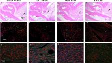
          Lyme disease is caused by Borrelia burgdorferi, and the pathogenesis of the disease is complex with both bacterial and host factors contributing to inflammatory responses. Lyme disease affects different organs including joints and results in arthritis. Immune responses stimulated by B. burgdorferi through toll-like receptors cause infiltration of leukocytes, which produce inflammatory cytokines and facilitate spirochete clearance. However, arthritic manifestations and chronic fatigue syndrome-like symptoms persist long after completion of antibiotic treatment regimens in a significant number of patients. To counter the effects of inflammation, treatment by non-steroidal anti-inflammatory drugs, hydroxychloroquine, or synovectomy to eradicate inflammatory arthritis in the involved joint could be employed; however, they often have long-term consequences. Acupuncture has been used for a long time in Asian medicine to diminish pain during various ailments, but the effects and its mechanism are just beginning to be explored. Control of inflammation by neuronal stimulation has been exploited as a systemic therapeutic intervention to arrest inflammatory processes. Our objective was to determine whether activation of the sciatic–vagal network by electroacupuncture on ST36 acupoint, which is used to control systemic inflammation in experimental models of infectious disorders such as endotoxemia, can also alleviate Lyme arthritis symptoms in mice. This aim was further strengthened by the reports that sciatic–vagal neuronal network stimulation can lead to dopamine production in the adrenal medulla and moderate the production of inflammatory factors. We first assessed whether electroacupuncture affects spirochete colonization to attenuate Lyme arthritis. Interestingly, bioluminescent B. burgdorferi burden detected by live imaging and qPCR were similar in electroacupuncture- and mock-treated mice, while electroacupuncture induced a lasting anti-inflammatory effect on mice. Despite the discontinuation of treatment at 2 weeks, the simultaneous decrease in neutrophils in the joints and inflammatory cytokine levels throughout the body at 4 weeks suggests a systemic and persistent effect of electroacupuncture that attenuates Lyme arthritis. Our results suggest that electroacupuncture-mediated anti-inflammatory responses could offer promising healthcare benefits in patients suffering from long-term Lyme disease manifestations.

          Related collections

          Most cited references64

          • Record: found
          • Abstract: found
          • Article: not found

          Nicotinic acetylcholine receptor alpha7 subunit is an essential regulator of inflammation.

          Excessive inflammation and tumour-necrosis factor (TNF) synthesis cause morbidity and mortality in diverse human diseases including endotoxaemia, sepsis, rheumatoid arthritis and inflammatory bowel disease. Highly conserved, endogenous mechanisms normally regulate the magnitude of innate immune responses and prevent excessive inflammation. The nervous system, through the vagus nerve, can inhibit significantly and rapidly the release of macrophage TNF, and attenuate systemic inflammatory responses. This physiological mechanism, termed the 'cholinergic anti-inflammatory pathway' has major implications in immunology and in therapeutics; however, the identity of the essential macrophage acetylcholine-mediated (cholinergic) receptor that responds to vagus nerve signals was previously unknown. Here we report that the nicotinic acetylcholine receptor alpha7 subunit is required for acetylcholine inhibition of macrophage TNF release. Electrical stimulation of the vagus nerve inhibits TNF synthesis in wild-type mice, but fails to inhibit TNF synthesis in alpha7-deficient mice. Thus, the nicotinic acetylcholine receptor alpha7 subunit is essential for inhibiting cytokine synthesis by the cholinergic anti-inflammatory pathway.
            Bookmark
            • Record: found
            • Abstract: found
            • Article: not found

            Preferential recruitment of CCR6-expressing Th17 cells to inflamed joints via CCL20 in rheumatoid arthritis and its animal model

            This report shows that interleukin (IL) 17–producing T helper type 17 (Th17) cells predominantly express CC chemokine receptor (CCR) 6 in an animal model of rheumatoid arthritis (RA). Th17 cells induced in vivo in normal mice via homeostatic proliferation similarly express CCR6, whereas those inducible in vitro by transforming growth factor β and IL-6 additionally need IL-1 and neutralization of interferon (IFN) γ and IL-4 for CCR6 expression. Forced expression of RORγt, a key transcription factor for Th17 cell differentiation, induces not only IL-17 but also CCR6 in naive T cells. Furthermore, Th17 cells produce CCL20, the known ligand for CCR6. Synoviocytes from arthritic joints of mice and humans also produce a large amount of CCL20, with a significant correlation (P = 0.014) between the amounts of IL-17 and CCL20 in RA joints. The CCL20 production by synoviocytes is augmented in vitro by IL-1β, IL-17, or tumor necrosis factor α, and is suppressed by IFN-γ or IL-4. Administration of blocking anti-CCR6 monoclonal antibody substantially inhibits mouse arthritis. Thus, the joint cytokine milieu formed by T cells and synovial cells controls the production of CCL20 and, consequently, the recruitment of CCR6+ arthritogenic Th17 cells to the inflamed joints. These results indicate that CCR6 expression contributes to Th17 cell function in autoimmune disease, especially in autoimmune arthritis such as RA.
              Bookmark
              • Record: found
              • Abstract: found
              • Article: not found

              The chemokine receptors CXCR3 and CCR5 mark subsets of T cells associated with certain inflammatory reactions.

              T cells infiltrating inflammatory sites are usually of the activated/memory type. The precise mechanism for the positioning of these cells within tissues is unclear. Adhesion molecules certainly play a role; however, the intricate control of cell migration appears to be mediated by numerous chemokines and their receptors. Particularly important chemokines for activated/memory T cells are the CXCR3 ligands IP-10 and Mig and the CCR5 ligands RANTES, macrophage inflammatory protein-1alpha, and macrophage inflammatory protein-1beta. We raised anti-CXCR3 mAbs and were able to detect high levels of CXCR3 expression on activated T cells. Surprisingly, a proportion of circulating blood T cells, B cells, and natural killer cells also expressed CXCR3. CCR5 showed a similar expression pattern as CXCR3, but was expressed on fewer circulating T cells. Blood T cells expressing CXCR3 (and CCR5) were mostly CD45RO+, and generally expressed high levels of beta1 integrins. This phenotype resembled that of T cells infiltrating inflammatory lesions. Immunostaining of T cells in rheumatoid arthritis synovial fluid confirmed that virtually all such T cells expressed CXCR3 and approximately 80% expressed CCR5, representing high enrichment over levels of CXCR3+ and CCR5+ T cells in blood, 35 and 15%, respectively. Analysis by immunohistochemistry of various inflamed tissues gave comparable findings in that virtually all T cells within the lesions expressed CXCR3, particularly in perivascular regions, whereas far fewer T cells within normal lymph nodes expressed CXCR3 or CCR5. These results demonstrate that the chemokine receptor CXCR3 and CCR5 are markers for T cells associated with certain inflammatory reactions, particularly TH-1 type reactions. Moreover, CXCR3 and CCR5 appear to identify subsets of T cells in blood with a predilection for homing to these sites.
                Bookmark

                Author and article information

                Contributors
                Journal
                Front Immunol
                Front Immunol
                Front. Immunol.
                Frontiers in Immunology
                Frontiers Media S.A.
                1664-3224
                18 July 2022
                2022
                : 13
                : 930287
                Affiliations
                [1] 1 Department of Microbiology, Biochemistry and Molecular Genetics, Rutgers New Jersey Medical School , Newark, NJ, United States
                [2] 2 Center of Perioperative Organ Protection, Department of Anesthesiology, Duke University , Durham, NC, United States
                Author notes

                Edited by: Fulvio D’Acquisto, University of Roehampton London, United Kingdom

                Reviewed by: Xu-Yun Hua, Shanghai University of Traditional Chinese Medicine, China; Jing Jiang, Beijing University of Chinese Medicine, China; Huirong Liu, Shanghai Research Center for Acupuncture and Meridians, China

                *Correspondence: Nikhat Parveen, Parveeni@ 123456njms.rutgers.edu

                †These authors have contributed equally to this work

                This article was submitted to Inflammation, a section of the journal Frontiers in Immunology

                Article
                10.3389/fimmu.2022.930287
                9342905
                35924250
                c3a970b1-09ec-40dc-9c50-44dd9081061a
                Copyright © 2022 Akoolo, Djokic, Rocha, Ulloa and Parveen

                This is an open-access article distributed under the terms of the Creative Commons Attribution License (CC BY). The use, distribution or reproduction in other forums is permitted, provided the original author(s) and the copyright owner(s) are credited and that the original publication in this journal is cited, in accordance with accepted academic practice. No use, distribution or reproduction is permitted which does not comply with these terms.

                History
                : 27 April 2022
                : 17 June 2022
                Page count
                Figures: 7, Tables: 0, Equations: 0, References: 64, Pages: 14, Words: 7176
                Funding
                Funded by: National Institutes of Health, doi 10.13039/100000002;
                Award ID: R01AI089921
                Categories
                Immunology
                Original Research

                Immunology
                borrelia burgdorferi,lyme arthritis,inflammation,vagus nerve,sciatic nerve,electroacupuncture

                Comments

                Comment on this article