3
views
0
recommends
+1 Recommend
0 collections
    0
    shares
      • Record: found
      • Abstract: found
      • Article: found
      Is Open Access

      Case report and review of literature: Isolated intramedullary spinal neurocysticercosis

      case-report

      Read this article at

      Bookmark
          There is no author summary for this article yet. Authors can add summaries to their articles on ScienceOpen to make them more accessible to a non-specialist audience.

          Abstract

          Background

          Cases of isolated intramedullary spinal neurocysticercosis are extremely rare. Only 25 cases have been reported before 2022. Due to its rarity, the diagnosis of spinal neurocysticercosis may be missed.

          Case presentation

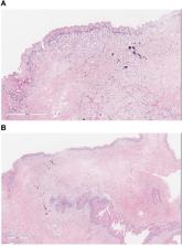
          We describe a 37-year-old female patient who developed back pain and lower extremity weakness and was found to have an intramedullary thoracic spine cystic lesion. She was taken to the operating room for resection of the lesion. Pathology revealed a larval cyst wall consistent with neurocysticercosis. The patient was started on albendazole and dexamethasone. Her exam improved post-operatively, and she was able to ambulate with minimal difficulty at the time of follow up.

          Conclusion

          The case provides insights on the diagnosis and treatment of isolated intramedullary spinal neurocysticercosis. Review of the literature suggests that combined surgical and medical intervention results in significant improvement in the patient's neurological exam, and decreases morbidity associated with the disease. We propose a treatment paradigm for this rare manifestation of neurocysticercosis.

          Related collections

          Most cited references25

          • Record: found
          • Abstract: found
          • Article: not found

          Taenia solium cysticercosis.

          The larval stage of the pork tapeworm (Taenia solium) infects the human nervous system, causing neurocysticercosis. This disease is one of the main causes of epileptic seizures in many less developed countries and is also increasingly seen in more developed countries because of immigration from endemic areas. Little information is available on the natural evolution of taeniasis or cysticercosis. Available therapeutic measures include steroids, treatments for symptoms, surgery, and, more controversially, antiparasitic drugs to kill brain parasites. Efforts to control and eliminate this disease are underway through antiparasitic treatment of endemic populations, development of pig vaccines, and other measures.
            Bookmark
            • Record: found
            • Abstract: found
            • Article: not found

            Clinical symptoms, diagnosis, and treatment of neurocysticercosis.

            The infection of the nervous system by the cystic larvae of Taenia solium (neurocysticercosis) is a frequent cause of seizure disorders. Neurocysticercosis is endemic or presumed to be endemic in many low-income countries. The lifecycle of the worm and the clinical manifestations of neurocysticercosis are well established, and CT and MRI have substantially improved knowledge of the disease course. Improvements in immunodiagnosis have further advanced comprehension of the pathophysiology of this disease. This knowledge has led to individualised treatment approaches that account for the involvement of parenchymal or extraparenchymal spaces, the number and form of parasites, and the extent of degeneration and associated inflammation. Clinical investigations are focused on development of effective treatments and reduction of side-effects induced by treatment, such as seizures, hydrocephalus, infarcts, and neuroinjury.
              Bookmark
              • Record: found
              • Abstract: found
              • Article: not found

              Diagnosis and treatment of neurocysticercosis.

              Neurocysticercosis is a parasitic disease caused by the larval (cystic) form of the pork cestode tapeworm, Taenia solium, and is a major cause of acquired seizures and epilepsy worldwide. Development of sensitive and specific diagnostic methods, particularly CT and MRI, has revolutionized our knowledge of the burden of cysticercosis infection and disease, and has led to the development of effective antihelminthic treatments for neurocysticercosis. The importance of calcified granulomas with perilesional edema as foci of seizures and epilepsy in populations where neurocysticercosis is endemic is newly recognized, and indicates that treatment with anti-inflammatory agents could have a role in controlling or preventing epilepsy in these patients. Importantly, neurocysticercosis is one of the few diseases that could potentially be controlled or eliminated-an accomplishment that would prevent millions of cases of epilepsy. This Review examines the rationale for treatment of neurocysticercosis and highlights the essential role of inflammation in the pathogenesis of disease, the exacerbation of symptoms that occurs as a result of antihelminthic treatment, and the limitations of current antihelminthic and anti-inflammatory treatments.
                Bookmark

                Author and article information

                Contributors
                Journal
                Front Neurol
                Front Neurol
                Front. Neurol.
                Frontiers in Neurology
                Frontiers Media S.A.
                1664-2295
                10 November 2022
                2022
                : 13
                : 1030468
                Affiliations
                [1] 1Department of Neurology, Loyola University Medical Center , Maywood, IL, United States
                [2] 2Department of Neurological Surgery, Loyola University Medical Center , Maywood, IL, United States
                [3] 3Department of Neurological Surgery, Riverside Medical Center , Kankakee, IL, United States
                [4] 4Department of Pathology and Laboratory Medicine, Loyola University Medical Center , Maywood, IL, United States
                [5] 5Department of Radiology, Loyola University Medical Center , Maywood, IL, United States
                Author notes

                Edited by: Alessia Libera Gazzonis, University of Milan, Italy

                Reviewed by: Sant Muangnoicharoen, Mahidol University, Thailand; Edvin Zekaj, Galeazzi Orthopedic Institute (IRCCS), Italy

                *Correspondence: John T. Tsiang jth.tsiang@ 123456gmail.com

                This article was submitted to Neuroinfectious Diseases, a section of the journal Frontiers in Neurology

                †These authors have contributed equally to this work

                Article
                10.3389/fneur.2022.1030468
                9687096
                36438967
                c3510ff3-696c-43b2-820a-c9e7916e93f9
                Copyright © 2022 Andino, Tsiang, Pecoraro, Jani, Iordanou, Zakaria, Borys, Pasquale, Nockels and Schneck.

                This is an open-access article distributed under the terms of the Creative Commons Attribution License (CC BY). The use, distribution or reproduction in other forums is permitted, provided the original author(s) and the copyright owner(s) are credited and that the original publication in this journal is cited, in accordance with accepted academic practice. No use, distribution or reproduction is permitted which does not comply with these terms.

                History
                : 29 August 2022
                : 21 October 2022
                Page count
                Figures: 5, Tables: 1, Equations: 0, References: 25, Pages: 7, Words: 3620
                Categories
                Neurology
                Case Report

                Neurology
                neurocysticercosis,spine,lesion,cyst,case report,resection,intramedullary
                Neurology
                neurocysticercosis, spine, lesion, cyst, case report, resection, intramedullary

                Comments

                Comment on this article