15
views
0
recommends
+1 Recommend
0 collections
    0
    shares
      • Record: found
      • Abstract: found
      • Article: found
      Is Open Access

      Brillouin microscopy: assessing ocular tissue biomechanics

      research-article

      Read this article at

      Bookmark
          There is no author summary for this article yet. Authors can add summaries to their articles on ScienceOpen to make them more accessible to a non-specialist audience.

          Abstract

          Purpose of review

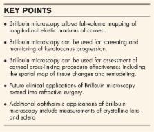
          Assessment of corneal biomechanics has been an unmet clinical need in ophthalmology for many years. Many researchers and clinicians have identified corneal biomechanics as source of variability in refractive procedures and one of the main factors in keratoconus. However, it has been difficult to accurately characterize corneal biomechanics in patients. The recent development of Brillouin light scattering microscopy heightens the promise of bringing biomechanics into the clinic. The aim of this review is to overview the progress and discuss prospective applications of this new technology.

          Recent findings

          Brillouin microscopy uses a low-power near-infrared laser beam to determine longitudinal modulus or mechanical compressibility of tissue by analyzing the return signal spectrum. Human clinical studies have demonstrated significant difference in the elastic properties of normal corneas versus corneas diagnosed with mild and severe keratoconus. Clinical data have also shown biomechanical changes after corneal cross-linking treatment of keratoconus patients. Brillouin measurements of the crystalline lens and sclera have also been demonstrated.

          Summary

          Brillouin microscopy is a promising technology under commercial development at present. The technique enables physicians to characterize the biomechanical properties of ocular tissues.

          Related collections

          Most cited references54

          • Record: found
          • Abstract: found
          • Article: not found

          Introduction of Two Novel Stiffness Parameters and Interpretation of Air Puff-Induced Biomechanical Deformation Parameters With a Dynamic Scheimpflug Analyzer.

          To investigate two new stiffness parameters and their relationships with the dynamic corneal response (DCR) parameters and compare normal and keratoconic eyes.
            Bookmark
            • Record: found
            • Abstract: found
            • Article: not found

            Corneal biomechanical properties in normal, post-laser in situ keratomileusis, and keratoconic eyes.

            To compare the biomechanical properties of normal, post-laser in situ keratomileusis (LASIK), and keratoconic corneas evaluated by corneal hysteresis and the corneal resistance factor measured with the Reichert Ocular Response Analyzer (ORA). Instituto Oftalmológico de Alicante, Vissum, Alicante, Spain. Two hundred fifty eyes were divided into 3 groups: normal (control group), post-LASIK, and keratoconus. The corneal biomechanical properties were measured with the ORA, which uses a dynamic bidirectional applanation process. The main outcome measures were intraocular pressure, corneal hysteresis, and the corneal resistance factor. The control group had 165 eyes; the LASIK group, 65 eyes; and the keratoconus group, 21 eyes. In the control group, the mean corneal hysteresis value was 10.8 mm Hg +/- 1.5 (SD) and the mean corneal resistance factor, 11.0 +/- 1.6 mm Hg. The corneal hysteresis value was lower in older eyes, and the difference between the youngest age group (9 to 14 years) and oldest age group (60 to 80 years) was statistically significant (P = .01, t test). One month after LASIK, corneal hysteresis and the corneal resistance factor decreased significantly, from 10.44 to 9.3 mm Hg and from 10.07 to 8.13 mm Hg, respectively. In the keratoconus group, the mean corneal hysteresis was 7.5 +/- 1.2 mm Hg and the mean corneal resistance factor, 6.2 +/- 1.9 mm Hg. There were statistically significant differences in both biomechanical parameters between keratoconic eyes and post-LASIK eyes (P<.001, t test). The corneal hysteresis and corneal resistance factor values were significantly lower in keratoconic eyes than in post-LASIK eyes. Future work is needed to determine whether these differences are useful in detecting keratoconus when other diagnostic tests are equivocal.
              Bookmark
              • Record: found
              • Abstract: found
              • Article: not found

              Assessment of the biomechanical properties of the cornea with the ocular response analyzer in normal and keratoconic eyes.

              To compare hysteresis, a novel measure of ocular rigidity (viscoelasticity) in normal and keratoconic eyes. The study consisted of 207 normal and 93 keratoconic eyes. Eyes were diagnosed as keratoconic based on clinical examination and corneal topography. The hysteresis was measured by the Ocular Response Analyzer (ORA; Reichert Ophthalmic Instruments, Buffalo, NY). The data were recorded by Generation 3 software for the ORA. Central corneal thickness (CCT) was measured with a handheld ultrasonic pachymeter in the midpupillary axis. The mean hysteresis was 10.7 +/- 2.0 (SD) mm Hg (range, 6.1-17.6) in normal eyes compared with 9.6 +/- 2.2 mm Hg (range, 4.7-16.7) in keratoconic eyes. The difference was statistically significant (P < 0.0001, unpaired t-test). Mean CCT in the normal and keratoconic eyes was 545.0 +/- 36.4 microm (range, 471-650) and 491.8 +/- 54.7 microm (range, 341-611), respectively; the difference was significant (P < 0.0001, unpaired t-test). Hysteresis was significantly higher in normal than in keratoconic eyes. It may be a useful measurement in addition to CCT, when assessing ocular rigidity, and may be of particular importance when trying to correct intraocular measurements for increased or decreased ocular rigidity. Long-term studies of change in hysteresis may provide information on the progression of keratoconus.
                Bookmark

                Author and article information

                Journal
                Curr Opin Ophthalmol
                Curr Opin Ophthalmol
                COOPH
                Current Opinion in Ophthalmology
                Lippincott Williams & Wilkins
                1040-8738
                1531-7021
                July 2018
                07 June 2018
                : 29
                : 4
                : 299-305
                Affiliations
                [a ]Harvard Medical School and Wellman Center for Photomedicine, Massachusetts General Hospital
                [b ]Intelon Optics Inc., Zero Emerson Place, Boston Massachusetts, USA
                Author notes
                Correspondence to Seok Hyun Yun, Professor and Patricia and Scott Eston MGH Research Scholar, Massachusetts General Hospital, 65 Landsdowne St. UP-525, Cambridge, MA 02139, USA. Tel: +1 617 768 8704; e-mail: syun@ 123456mgh.harvard.edu
                Article
                ICU290405 00006
                10.1097/ICU.0000000000000489
                6012042
                29771749
                c1dd715b-8de3-4ff3-8c34-c49141ae1b3b
                Copyright © 2018 The Author(s). Published by Wolters Kluwer Health, Inc.

                This is an open access article distributed under the terms of the Creative Commons Attribution-Non Commercial-No Derivatives License 4.0 (CCBY-NC-ND), where it is permissible to download and share the work provided it is properly cited. The work cannot be changed in any way or used commercially without permission from the journal. http://creativecommons.org/licenses/by-nc-nd/4.0

                History
                Categories
                REFRACTIVE SURGERY: Edited by Jimmy K. Lee
                Custom metadata
                TRUE

                biomechanics,brillouin spectroscopy,cornea,keratoconus,refractive surgery

                Comments

                Comment on this article