0
views
0
recommends
+1 Recommend
0 collections
    0
    shares
      • Record: found
      • Abstract: found
      • Article: found
      Is Open Access

      Cisplatin ototoxicity mechanism and antagonistic intervention strategy: a scope review

      review-article

      Read this article at

      Bookmark
          There is no author summary for this article yet. Authors can add summaries to their articles on ScienceOpen to make them more accessible to a non-specialist audience.

          Abstract

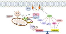
          Cisplatin is a first-line chemotherapeutic agent in the treatment of malignant tumors with remarkable clinical effects and low cost. However, the ototoxicity and neurotoxicity of cisplatin greatly limit its clinical application. This article reviews the possible pathways and molecular mechanisms of cisplatin trafficking from peripheral blood into the inner ear, the toxic response of cisplatin to inner ear cells, as well as the cascade reactions leading to cell death. Moreover, this article highlights the latest research progress in cisplatin resistance mechanism and cisplatin ototoxicity. Two effective protective mechanisms, anti-apoptosis and mitophagy activation, and their interaction in the inner ear are discussed. Additionally, the current clinical preventive measures and novel therapeutic agents for cisplatin ototoxicity are described. Finally, this article also forecasts the prospect of possible drug targets for mitigating cisplatin-induced ototoxicity. These include the use of antioxidants, inhibitors of transporter proteins, inhibitors of cellular pathways, combination drug delivery methods, and other mechanisms that have shown promise in preclinical studies. Further research is needed to evaluate the efficacy and safety of these approaches.

          Related collections

          Most cited references89

          • Record: found
          • Abstract: found
          • Article: not found

          Ubiquitin is phosphorylated by PINK1 to activate parkin.

          PINK1 (PTEN induced putative kinase 1) and PARKIN (also known as PARK2) have been identified as the causal genes responsible for hereditary recessive early-onset Parkinsonism. PINK1 is a Ser/Thr kinase that specifically accumulates on depolarized mitochondria, whereas parkin is an E3 ubiquitin ligase that catalyses ubiquitin transfer to mitochondrial substrates. PINK1 acts as an upstream factor for parkin and is essential both for the activation of latent E3 parkin activity and for recruiting parkin onto depolarized mitochondria. Recently, mechanistic insights into mitochondrial quality control mediated by PINK1 and parkin have been revealed, and PINK1-dependent phosphorylation of parkin has been reported. However, the requirement of PINK1 for parkin activation was not bypassed by phosphomimetic parkin mutation, and how PINK1 accelerates the E3 activity of parkin on damaged mitochondria is still obscure. Here we report that ubiquitin is the genuine substrate of PINK1. PINK1 phosphorylated ubiquitin at Ser 65 both in vitro and in cells, and a Ser 65 phosphopeptide derived from endogenous ubiquitin was only detected in cells in the presence of PINK1 and following a decrease in mitochondrial membrane potential. Unexpectedly, phosphomimetic ubiquitin bypassed PINK1-dependent activation of a phosphomimetic parkin mutant in cells. Furthermore, phosphomimetic ubiquitin accelerates discharge of the thioester conjugate formed by UBCH7 (also known as UBE2L3) and ubiquitin (UBCH7∼ubiquitin) in the presence of parkin in vitro, indicating that it acts allosterically. The phosphorylation-dependent interaction between ubiquitin and parkin suggests that phosphorylated ubiquitin unlocks autoinhibition of the catalytic cysteine. Our results show that PINK1-dependent phosphorylation of both parkin and ubiquitin is sufficient for full activation of parkin E3 activity. These findings demonstrate that phosphorylated ubiquitin is a parkin activator.
            Bookmark
            • Record: found
            • Abstract: found
            • Article: found
            Is Open Access

            Cisplatin Induces a Mitochondrial-ROS Response That Contributes to Cytotoxicity Depending on Mitochondrial Redox Status and Bioenergetic Functions

            Cisplatin is one of the most effective and widely used anticancer agents for the treatment of several types of tumors. The cytotoxic effect of cisplatin is thought to be mediated primarily by the generation of nuclear DNA adducts, which, if not repaired, cause cell death as a consequence of DNA replication and transcription blockage. However, the ability of cisplatin to induce nuclear DNA (nDNA) damage per se is not sufficient to explain its high degree of effectiveness nor the toxic effects exerted on normal, post-mitotic tissues. Oxidative damage has been observed in vivo following exposure to cisplatin in several tissues, suggesting a role for oxidative stress in the pathogenesis of cisplatin-induced dose-limiting toxicities. However, the mechanism of cisplatin-induced generation of ROS and their contribution to cisplatin cytotoxicity in normal and cancer cells is still poorly understood. By employing a panel of normal and cancer cell lines and the budding yeast Saccharomyces cerevisiae as model system, we show that exposure to cisplatin induces a mitochondrial-dependent ROS response that significantly enhances the cytotoxic effect caused by nDNA damage. ROS generation is independent of the amount of cisplatin-induced nDNA damage and occurs in mitochondria as a consequence of protein synthesis impairment. The contribution of cisplatin-induced mitochondrial dysfunction in determining its cytotoxic effect varies among cells and depends on mitochondrial redox status, mitochondrial DNA integrity and bioenergetic function. Thus, by manipulating these cellular parameters, we were able to enhance cisplatin cytotoxicity in cancer cells. This study provides a new mechanistic insight into cisplatin-induced cell killing and may lead to the design of novel therapeutic strategies to improve anticancer drug efficacy.
              Bookmark
              • Record: found
              • Abstract: found
              • Article: found
              Is Open Access

              Autophagy protects auditory hair cells against neomycin-induced damage

              ABSTRACT Aminoglycosides are toxic to sensory hair cells (HCs). Macroautophagy/autophagy is an essential and highly conserved self-digestion pathway that plays important roles in the maintenance of cellular function and viability under stress. However, the role of autophagy in aminoglycoside-induced HC injury is unknown. Here, we first found that autophagy activity was significantly increased, including enhanced autophagosome-lysosome fusion, in both cochlear HCs and HEI-OC-1 cells after neomycin or gentamicin injury, suggesting that autophagy might be correlated with aminoglycoside-induced cell death. We then used rapamycin, an autophagy activator, to increase the autophagy activity and found that the ROS levels, apoptosis, and cell death were significantly decreased after neomycin or gentamicin injury. In contrast, treatment with the autophagy inhibitor 3-methyladenine (3-MA) or knockdown of autophagy-related (ATG) proteins resulted in reduced autophagy activity and significantly increased ROS levels, apoptosis, and cell death after neomycin or gentamicin injury. Finally, after neomycin injury, the antioxidant N-acetylcysteine could successfully prevent the increased apoptosis and HC loss induced by 3-MA treatment or ATG knockdown, suggesting that autophagy protects against neomycin-induced HC damage by inhibiting oxidative stress. We also found that the dysfunctional mitochondria were not eliminated by selective autophagy (mitophagy) in HEI-OC-1 cells after neomycin treatment, suggesting that autophagy might not directly target the damaged mitochondria for degradation. This study demonstrates that moderate ROS levels can promote autophagy to recycle damaged cellular constituents and maintain cellular homeostasis, while the induction of autophagy can inhibit apoptosis and protect the HCs by suppressing ROS accumulation after aminoglycoside injury.
                Bookmark

                Author and article information

                Contributors
                Journal
                Front Cell Neurosci
                Front Cell Neurosci
                Front. Cell. Neurosci.
                Frontiers in Cellular Neuroscience
                Frontiers Media S.A.
                1662-5102
                01 June 2023
                2023
                : 17
                : 1197051
                Affiliations
                [1] 1Department of Otorhinolaryngology–Head and Neck Surgery, School of Medicine, Xinhua Hospital, Shanghai Jiao Tong University , Shanghai, China
                [2] 2School of Medicine, Ear Institute, Shanghai Jiao Tong University , Shanghai, China
                [3] 3Key Laboratory of Translational Medicine on Ear and Nose Diseases , Shanghai, China
                Author notes

                Edited by: Zuhong He, Wuhan University, China

                Reviewed by: Fuping Qian, Nantong University, China; Benjamin Philipp Ernst, University Hospital Bonn, Germany

                *Correspondence: Guiliang Zheng, icing_blue@ 123456126.comm
                Article
                10.3389/fncel.2023.1197051
                10267334
                37323582
                c197154c-1eb8-4aee-9564-38916ae91284
                Copyright © 2023 Li, Zhang, Song, Gao, Li, Jie, Huang, Zheng, Yang and He.

                This is an open-access article distributed under the terms of the Creative Commons Attribution License (CC BY). The use, distribution or reproduction in other forums is permitted, provided the original author(s) and the copyright owner(s) are credited and that the original publication in this journal is cited, in accordance with accepted academic practice. No use, distribution or reproduction is permitted which does not comply with these terms.

                History
                : 30 March 2023
                : 15 May 2023
                Page count
                Figures: 1, Tables: 4, Equations: 0, References: 89, Pages: 12, Words: 9938
                Funding
                Funded by: National Natural Science Foundation of China, doi 10.13039/501100001809;
                Award ID: No. 82271164
                Award ID: No. 1970881
                Award ID: No. 82071069
                This work was supported by the National Natural Science Foundation of China (Nos. 82271164, 81970881, and 82071069).
                Categories
                Neuroscience
                Review
                Custom metadata
                Cellular Neuropathology

                Neurosciences
                cisplatin,ototoxicity,apoptosis,mitophagy,antioxidants
                Neurosciences
                cisplatin, ototoxicity, apoptosis, mitophagy, antioxidants

                Comments

                Comment on this article