10
views
0
recommends
+1 Recommend
0 collections
    0
    shares
      • Record: found
      • Abstract: found
      • Article: found
      Is Open Access

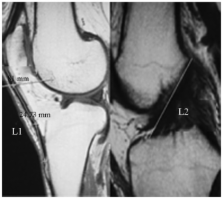
      Measurement of normal patellar ligament and anterior cruciate ligament by MRI and data analysis

      research-article

      Read this article at

      Bookmark
          There is no author summary for this article yet. Authors can add summaries to their articles on ScienceOpen to make them more accessible to a non-specialist audience.

          Abstract

          The aim of this study was to obtain geometric data of in vivo patellar ligament (PL) and anterior cruciate ligament (ACL) by MRI and to analyze the correlation of the two with body weight, height and gender. A total of 157 cases with normal sagittal images of bilateral PL and ACL were enrolled. The PL and ACL lengths in the images were measured using the Radworks 5.1 application. The intraclass correlation coefficient for the data measured independently by three doctors was 0.997–1.000. In individuals aged 15–24 years, the values of PL and ACL length and the PL to ACL ratio were 43.95±4.25 mm, 38.45±4.62 mm and 1.15±1.09 in males and 42.03±0.94 mm, 36.00±1.06 mm and 1.18±0.1 in females, respectively. In individuals aged 25–64 years, the values in males were 40.99±4.45 mm, 36.06±3.74 mm and 1.14±0.09 and in females were 39.84±0.64 mm, 36.50±0.81 mm and 1.11±0.02, respectively. In individuals aged ≥65 years, the values in males were 41.43±3.08 mm, 36.62±3.44 mm and 1.15±0.09 and in females were 38.94±0.79 mm, 34.36±0.85 mm and 1.13±0.07, respectively. There was a significant difference between PL and ACL length on the same side (P<0.01). The data obtained was stable and repeatable. The present study established a database of PL and ACL length and the ratio of the two measured by MRI.

          Related collections

          Most cited references32

          • Record: found
          • Abstract: found
          • Article: not found

          Biomechanical analysis of human ligament grafts used in knee-ligament repairs and reconstructions.

          Virtually all types of collagenous tissues have been transferred in and around the knee joint for intra-articular and extra-articular ligament reconstructions. However, the mechanical properties (in particular, strength) of such grafts have not been determined in tissues from young adult donors, where age and disuse-related effects have been excluded. To provide this information, we subjected ligament graft tissues to high-strain-rate failure tests to determine their strength and elongation properties. The results were compared with the mechanical properties of anterior cruciate ligaments from a similar young-adult donor population. The study indicated that some graft tissues used in ligament reconstructions are markedly weak and therefore are at risk for elongation and failure at low forces. Grafts utilizing prepatellar retinacular tissues (as in certain anterior-cruciate reconstructions) and others in which a somewhat narrow width of fascia lata or distal iliotibial tract is utilized are included in this at-risk group. Wider grafts from the iliotibial tract or fascia lata would of course proportionally increase ultimate strength. The semitendinosus and gracilis tendons are stronger, having 70 and 49 per cent, respectively, of the initial strength of anterior cruciate ligaments. The bone-patellar tendon-bone graft (fourteen to fifteen millimeters wide, medial or central portion) was the strongest, with a mean strength of 159 to 168 per cent of that of anterior cruciate ligaments. Patellar tendon-bone units, based on grip-to-grip motions, were found to be three to four times stiffer than similarly gripped anterior cruciate ligaments, while gracilis and semitendinosus tendon preparations had values that were nearly identical to those of anterior cruciate ligaments.(ABSTRACT TRUNCATED AT 250 WORDS)
            Bookmark
            • Record: found
            • Abstract: found
            • Article: not found

            Ligament length relationships in the moving knee.

            This article presents an investigation of potential ligament attachment sites for surgical reconstruction of the anterior and posterior cruciate ligaments as well as for the lateral extraarticular iliotibial band tenodesis. Our methodology was based on quantitative measurements of knee anatomy and motion in fresh cadavers, not on biomechanical modeling. Using computer search techniques, we located all the ligament insertion sites that were nearly isometric for motion of the intact knee.
              Bookmark
              • Record: found
              • Abstract: found
              • Article: not found

              The geometry of patella and patellar tendon measured on knee MRI.

              This study provides the geometry of patella and patellar tendon measured on sagittal and axial magnetic resonance images of 172 knees (142 males, 30 females) of 163 subjects (135 males, 28 females) whose mean age was 26.7. The gender difference and the relationship with anthropometry were also given. As for patella, the longitudinal lengths of the whole and articulating surface were 44.6, 32.9 mm, and the thickness was 22.3 mm. The mediolateral width was 45.8 mm, and the central ridge was located 19.9 mm or 43% lateral from the medial border. As for patellar tendon, the longitudinal length was 40.2 mm. The widths of proximal and distal part were 30.3, and 24.0 mm. The thicknesses of proximal and distal part were 3.2, and 5.0 mm. The geometry of the patella and patellar tendon was larger in male than in female (P < 0.001). Anthropometry including weight, height, body mass index corresponds well with the thickness of patella but poorly with the length of patellar tendon. These data can provide useful information in the field of knee surgery and sports medicine.
                Bookmark

                Author and article information

                Journal
                Exp Ther Med
                Exp Ther Med
                ETM
                Experimental and Therapeutic Medicine
                D.A. Spandidos
                1792-0981
                1792-1015
                March 2013
                17 January 2013
                17 January 2013
                : 5
                : 3
                : 917-921
                Affiliations
                [1 ]Departments of Radiology, The First Affiliated Hospital of Xinxiang Medical University, Henan, Weihui 453100, P.R. China
                [2 ]Gynecology and Obstetrics, The First Affiliated Hospital of Xinxiang Medical University, Henan, Weihui 453100, P.R. China
                Author notes
                Correspondence to: Professor Ruimin Yang, Department of Radiology, The First Affiliated Hospital of Xinxiang Medical University, 88 Jiankang Road, Henan, Weihui 453100, P.R. China, E-mail: ruiminyangcn@ 123456126.com
                Article
                etm-05-03-0917
                10.3892/etm.2013.906
                3570255
                bf5273ac-2f67-4ab7-bc8d-fd7ad696d0d2
                Copyright © 2013, Spandidos Publications

                This is an open-access article licensed under a Creative Commons Attribution-NonCommercial 3.0 Unported License. The article may be redistributed, reproduced, and reused for non-commercial purposes, provided the original source is properly cited.

                History
                : 19 October 2012
                : 20 December 2012
                Categories
                Articles

                Medicine
                anterior cruciate ligament,clinical anatomy,magnetic resonance imaging,patellar tendon

                Comments

                Comment on this article