3
views
0
recommends
+1 Recommend
0 collections
    0
    shares
      • Record: found
      • Abstract: found
      • Article: found
      Is Open Access

      Morphological and immunohistochemical characterization of spontaneous endometriosis in rhesus macaques ( Macaca mulatta)

      research-article
      , ,
      Primate Biology
      Copernicus GmbH

      Read this article at

      Bookmark
          There is no author summary for this article yet. Authors can add summaries to their articles on ScienceOpen to make them more accessible to a non-specialist audience.

          Abstract

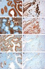
          Several cases of spontaneous endometriosis in middle-aged to old rhesus macaques ( Macaca mulatta) from the breeding colony of the German Primate Center were thoroughly characterized with regards to anatomical distribution and macroscopic appearance, histological differentiation and immunohistochemical profile including somatic markers, hormonal receptors, and proliferation indices. More than half of the examined animals (five of nine) were directly related to one breeding male, supporting a strong genetic predisposition. Histologically, four different types of endometriotic lesions, depending on the degree of ectopic endometrial gland and stromal differentiation (well differentiated, purely stromal, mixed differentiation, poorly differentiated), could be constantly identified within all animals. Immunohistochemistry (IHC) of cytokeratin (CK), vimentin, smooth muscle actin (SMA), desmin, estrogen (ER), and progesterone (PR) receptors as well as of the nuclear proteins Ki67 and p53 revealed varying staining patterns in the four different types of endometriosis differentiation and compared to normal endometrium. Purely stromal, mixed, or poorly differentiated lesions, especially, showed additional cytokeratin-positive stromal cells, whereas epithelial cells of endometriosis with mixed or poor differentiation increasingly expressed mesenchymal markers (vimentin, SMA). Hormonal receptor and Ki67 expression in well-differentiated endometriotic lesions mostly reflected that of normal endometrial tissue according to the cyclic phase of the animal, while the expression gradually diminished with decreasing grade of differentiation. However, increased nuclear accumulations of p53 antigen could only be continuously detected in epithelial cells of mixed or poorly differentiated endometriosis. Altogether, these findings support the pathogenetic theory of coelomic metaplasia, since the expression profiles of somatic markers in less differentiated forms closely resembled that of mesothelial cells. Thus, the four different histological types of endometriosis might display subsequent grades of differentiation in the course of time, with poorly differentiated types representing newly formed, immature lesions and well-differentiated types being older, fully differentiated forms, rather than being the outcome of dedifferentiation processes.

          Related collections

          Most cited references81

          • Record: found
          • Abstract: found
          • Article: found
          Is Open Access

          Endometrial stem/progenitor cells: the first 10 years

          BACKGROUND The existence of stem/progenitor cells in the endometrium was postulated many years ago, but the first functional evidence was only published in 2004. The identification of rare epithelial and stromal populations of clonogenic cells in human endometrium has opened an active area of research on endometrial stem/progenitor cells in the subsequent 10 years. METHODS The published literature was searched using the PubMed database with the search terms ‘endometrial stem cells and menstrual blood stem cells' until December 2014. RESULTS Endometrial epithelial stem/progenitor cells have been identified as clonogenic cells in human and as label-retaining or CD44+ cells in mouse endometrium, but their characterization has been modest. In contrast, endometrial mesenchymal stem/stromal cells (MSCs) have been well characterized and show similar properties to bone marrow MSCs. Specific markers for their enrichment have been identified, CD146+PDGFRβ+ (platelet-derived growth factor receptor beta) and SUSD2+ (sushi domain containing-2), which detected their perivascular location and likely pericyte identity in endometrial basalis and functionalis vessels. Transcriptomics and secretomics of SUSD2+ cells confirm their perivascular phenotype. Stromal fibroblasts cultured from endometrial tissue or menstrual blood also have some MSC characteristics and demonstrate broad multilineage differentiation potential for mesodermal, endodermal and ectodermal lineages, indicating their plasticity. Side population (SP) cells are a mixed population, although predominantly vascular cells, which exhibit adult stem cell properties, including tissue reconstitution. There is some evidence that bone marrow cells contribute a small population of endometrial epithelial and stromal cells. The discovery of specific markers for endometrial stem/progenitor cells has enabled the examination of their role in endometrial proliferative disorders, including endometriosis, adenomyosis and Asherman's syndrome. Endometrial MSCs (eMSCs) and menstrual blood stromal fibroblasts are an attractive source of MSCs for regenerative medicine because of their relative ease of acquisition with minimal morbidity. Their homologous and non-homologous use as autologous and allogeneic cells for therapeutic purposes is currently being assessed in preclinical animal models of pelvic organ prolapse and phase I/II clinical trials for cardiac failure. eMSCs and stromal fibroblasts also exhibit non-stem cell-associated immunomodulatory and anti-inflammatory properties, further emphasizing their desirable properties for cell-based therapies. CONCLUSIONS Much has been learnt about endometrial stem/progenitor cells in the 10 years since their discovery, although several unresolved issues remain. These include rationalizing the terminology and diagnostic characteristics used for distinguishing perivascular stem/progenitor cells from stromal fibroblasts, which also have considerable differentiation potential. The hierarchical relationship between clonogenic epithelial progenitor cells, endometrial and decidual SP cells, CD146+PDGFR-β+ and SUSD2+ cells and menstrual blood stromal fibroblasts still needs to be resolved. Developing more genetic animal models for investigating the role of endometrial stem/progenitor cells in endometrial disorders is required, as well as elucidating which bone marrow cells contribute to endometrial tissue. Deep sequencing and epigenetic profiling of enriched populations of endometrial stem/progenitor cells and their differentiated progeny at the population and single-cell level will shed new light on the regulation and function of endometrial stem/progenitor cells.
            Bookmark
            • Record: found
            • Abstract: found
            • Article: not found

            Role of K-ras and Pten in the development of mouse models of endometriosis and endometrioid ovarian cancer.

            Epithelial ovarian tumors present a complex clinical, diagnostic and therapeutic challenge because of the difficulty of early detection, lack of known precursor lesions and high mortality rates. Endometrioid ovarian carcinomas are frequently associated with endometriosis, but the mechanism for this association remains unknown. Here we present the first genetic models of peritoneal endometriosis and endometrioid ovarian adenocarcinoma in mice, both based on the activation of an oncogenic K-ras allele. In addition, we find that expression of oncogenic K-ras or conditional Pten deletion within the ovarian surface epithelium gives rise to preneoplastic ovarian lesions with an endometrioid glandular morphology. Furthermore, the combination of the two mutations in the ovary leads to the induction of invasive and widely metastatic endometrioid ovarian adenocarcinomas with complete penetrance and a disease latency of only 7 weeks. The ovarian cancer model described in this study recapitulates the specific tumor histomorphology and metastatic potential of the human disease.
              Bookmark
              • Record: found
              • Abstract: found
              • Article: found
              Is Open Access

              Uncovering the role of p53 splice variants in human malignancy: a clinical perspective

              Thirty-five years of research on p53 gave rise to more than 68,000 articles and reviews, but did not allow the uncovering of all the mysteries that this major tumor suppressor holds. How p53 handles the different signals to decide the appropriate cell fate in response to a stress and its implication in tumorigenesis and cancer progression remains unclear. Nevertheless, the uncovering of p53 isoforms has opened new perspectives in the cancer research field. Indeed, the human TP53 gene encodes not only one but at least twelve p53 protein isoforms, which are produced in normal tissues through alternative initiation of translation, usage of alternative promoters, and alternative splicing. In recent years, it became obvious that the different p53 isoforms play an important role in regulating cell fate in response to different stresses in normal cells by differentially regulating gene expression. In cancer cells, abnormal expression of p53 isoforms contributes actively to cancer formation and progression, regardless of TP53 mutation status. They can also be associated with response to treatment, depending on the cell context. The determination of p53 isoform expression and p53 mutation status helps to define different subtypes within a particular cancer type, which would have different responses to treatment. Thus, the understanding of the regulation of p53 isoform expression and their biological activities in relation to the cellular context would constitute an important step toward the improvement of the diagnostic, prognostic, and predictive values of p53 in cancer treatment. This review aims to summarize the involvement of p53 isoforms in cancer and to highlight novel potential therapeutic targets.
                Bookmark

                Author and article information

                Contributors
                Journal
                Primate Biol
                Primate Biol
                PB
                Primate Biology
                Copernicus GmbH
                2363-4707
                2363-4715
                13 April 2017
                2017
                : 4
                : 1
                : 77-91
                Affiliations
                [1]Pathology Unit, German Primate Center, 37077 Göttingen, Germany
                Author notes
                [*] Correspondence: Eva Gruber-Dujardin ( edujardin@ 123456dpz.eu )
                Article
                01021829
                10.5194/pb-4-77-2017
                7041517
                bf51dd04-3404-4611-89e1-1c7da60daff8
                Copyright: © 2017 Eva Gruber-Dujardin et al.

                This work is licensed under the Creative Commons Attribution 3.0 Unported License. To view a copy of this licence, visit https://creativecommons.org/licenses/by/3.0/

                History
                : 14 December 2016
                : 21 March 2017
                Categories
                Research Article

                Comments

                Comment on this article