2
views
0
recommends
+1 Recommend
0 collections
    0
    shares
      • Record: found
      • Abstract: found
      • Article: found
      Is Open Access

      Intracapsular Micro-Enucleation of a Painful Superficial Peroneal Nerve Schwannoma in a 60-Year Old Man: A Rare Encounter

      research-article

      Read this article at

      Bookmark
          There is no author summary for this article yet. Authors can add summaries to their articles on ScienceOpen to make them more accessible to a non-specialist audience.

          Abstract

          Patient: Male, 60-year-old

          Final Diagnosis: Right superficial peroneal nerve schwannoma

          Symptoms: Lump on the lateral aspect of the right upper leg • pain

          Medication: —

          Clinical Procedure: Intracapsular micro-enucleation of the lesion

          Specialty: Neurosurgery

          Objective:

          Rare disease

          Background:

          Schwannomas are the most common benign peripheral nerve sheath tumors, localized mainly to the cranial and upper extremity nerves. Their occurrence in the lower limbs is uncommon, and specific involvement of the superficial peroneal nerve is exceedingly rare. We report a case of a painful right superficial peroneal nerve schwannoma that was excised via the intracapsular micro-enucleation technique.

          Case Report:

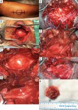
          A 60-year-old South Asian man presented with a 2-year history of a painful lump on the lateral aspect of the right upper leg. Clinical examination revealed a firm mass located at the proximal lateral aspect of the right leg, measuring approximately 3×2.5 cm. Severe tenderness over the mass was present. The Tinel test was positive. There were no sensory or motor deficits or history of neurofibromatosis. Imaging showed features suggestive of a schwannoma. Surgery was indicated; intracapsular micro-enucleation was performed. Histopathological assessment of the tumor demonstrated Antoni A and B patterns with nuclear palisading and Verocay bodies, hallmarks of a schwannoma. The postoperative period was uneventful; no neurological deficits were noted.

          Conclusions:

          The case described is considered rare, with no data on disease epidemiology in the literature. We provide a brief review and add pivotal data to the literature. Despite its rarity, one should remain cognizant of the condition and consider it in the differential diagnosis of nontraumatic leg pain. Based on our experience, corroboration from previous case reports, and the satisfactory outcome of our case, we advocate the intracapsular micro-enucleation technique when possible for schwannomas.

          Related collections

          Most cited references32

          • Record: found
          • Abstract: found
          • Article: not found

          Pathology of peripheral nerve sheath tumors: diagnostic overview and update on selected diagnostic problems.

          Peripheral nerve sheath tumors are common neoplasms, with classic identifiable features, but on occasion, they are diagnostically challenging. Although well-defined subtypes of peripheral nerve sheath tumors were described early in the history of surgical pathology, controversies regarding the classification and grading of these tumors persist. Advances in molecular biology have provided new insights into the nature of the various peripheral nerve sheath tumors, and have begun to suggest novel targeted therapeutic approaches. In this review, we discuss current concepts and problematic areas in the pathology of peripheral nerve sheath tumors. Diagnostic criteria and differential diagnosis for the major categories of nerve sheath tumors are proposed, including neurofibroma, schwannoma, and perineurioma. Diagnostically challenging variants, including plexiform, cellular and melanotic schwannomas are highlighted. A subset of these affects the childhood population, and has historically been interpreted as malignant, although current evidence and outcome data suggest they represent benign entities. The growing current literature and the author's experience with difficult to classify borderline or "hybrid tumors" are discussed and illustrated. Some of these classification gray zones occur with frequency in the gastrointestinal tract, an anatomical compartment that must always be entertained when examining these neoplasms. Other growing recent areas of interest include the heterogeneous group of pseudoneoplastic lesions involving peripheral nerve composed of mature adipose tissue and/or skeletal muscle, such as the enigmatic neuromuscular choristoma. Malignant peripheral nerve sheath tumors (MPNST) represent a diagnostically controversial group; difficulties in grading and guidelines to separate "atypical neurofibroma" from MPNST are provided. There is an increasing literature of MPNST mimics which neuropathologists must be aware of, including synovial sarcoma and ossifying fibromyxoid tumor. Finally, we discuss entities that are lacking from the section on cranial and paraspinal nerves in the current WHO classification, and that may warrant inclusion in future classifications. In summary, although the diagnosis and classification of most conventional peripheral nerve sheath tumors are relatively straightforward for the experienced observer, yet borderline and difficult-to-classify neoplasms continue to be problematic. In the current review, we attempt to provide some useful guidelines for the surgical neuropathologist to help navigate these persistent, challenging problems.
            Bookmark
            • Record: found
            • Abstract: found
            • Article: not found

            From the archives of the AFIP. Imaging of musculoskeletal neurogenic tumors: radiologic-pathologic correlation.

            Numerous neurogenic tumors can affect the musculoskeletal system, including traumatic neuroma, Morton neuroma, neural fibrolipoma, nerve sheath ganglion, neurilemoma, neurofibroma, and malignant peripheral nerve sheath tumors (PNSTs). The diagnosis of neurogenic tumors can be suggested from their imaging appearances, including lesion shape and intrinsic imaging characteristics. It is also important to establish lesion location along a typical nerve distribution (eg, plantar digital nerve in Morton neuroma, median nerve in neural fibrolipoma, large nerve trunk in benign and malignant PNSTs). Traumatic and Morton neuromas are commonly related to an amputation stump or are located in the intermetatarsal space, respectively. Neural fibrolipomas show fat interspersed between nerve fascicles and are often associated with macrodactyly. Nerve sheath ganglion has a cystic appearance and commonly occurs about the knee. Radiologic characteristics of neurilemoma, neurofibroma, and malignant PNST at computed tomography (CT), ultrasonography, and magnetic resonance imaging include fusiform shape, identification of entering and exiting nerve, low attenuation at CT, target sign, fascicular sign, split-fat sign, and associated muscle atrophy. Although differentiation of neurilemoma from neurofibroma and of benign from malignant PNST is problematic, recognition of the radiologic appearances of neurogenic tumors often allows prospective diagnosis and improves clinical management of patients.
              Bookmark
              • Record: found
              • Abstract: found
              • Article: not found

              Benign solitary schwannomas: a review of 234 cases.

              We reviewed 234 benign solitary schwannomas treated between 1984 and 2004. The mean age of the patients was 45.2 years (11 to 82). There were 170 tumours (73%) in the upper limb, of which 94 (40%) arose from the brachial plexus or other nerves within the posterior triangle of the neck. Six (2.6%) were located within muscle or bone. Four patients (1.7%) presented with tetraparesis due to an intraspinal extension. There were 198 primary referrals (19 of whom had a needle biopsy in the referring unit) and in these patients the tumour was excised. After having surgery or an open biopsy at another hospital, a further 36 patients were seen because of increased neurological deficit, pain or incomplete excision. In these, a nerve repair was performed in 18 and treatment for pain or paralysis was offered to another 14. A tender mass was found in 194 (98%) of the primary referrals. A Tinel-like sign was recorded in 155 (81%). Persistent spontaneous pain occurred in 60 (31%) of the 194 with tender mass, impairment of cutaneous sensibility in 39 (20%), and muscle weakness in 24 (12%). After apparently adequate excision, two tumours recurred. No case of malignant transformation was seen.
                Bookmark

                Author and article information

                Journal
                Am J Case Rep
                Am J Case Rep
                amjcaserep
                The American Journal of Case Reports
                International Scientific Literature, Inc.
                1941-5923
                2022
                24 February 2022
                : 23
                : e936056-1-e936056-7
                Affiliations
                [1 ]Department of Neurosurgery, Ibn Sina Hospital, Ministry of Health, Kuwait City,Kuwait
                [2 ]Department of Histopathology, Sabah Hospital, Ministry of Health, Kuwait City, Kuwait
                Author notes
                Corresponding Author: Yousef A.I. Abousedu, e-mail: dr.abousedu@ 123456hotmail.com

                Authors’ Contribution:

                [A]

                Study Design

                [B]

                Data Collection

                [C]

                Statistical Analysis

                [D]

                Data Interpretation

                [E]

                Manuscript Preparation

                [F]

                Literature Search

                [G]

                Funds Collection

                Financial support: None declared

                Conflict of interest: None declared

                Author information
                https://orcid.org/0000-0002-2365-0132
                Article
                936056
                10.12659/AJCR.936056
                8886513
                35197439
                bf003bf1-7a68-429d-8beb-7f08620145ff
                © Am J Case Rep, 2022

                This work is licensed under Creative Common Attribution-NonCommercial-NoDerivatives 4.0 International ( CC BY-NC-ND 4.0)

                History
                : 10 January 2022
                : 07 February 2022
                : 15 February 2022
                Categories
                Articles

                middle east,neurilemmoma,peripheral nervous system neoplasms,peroneal nerve,schwannomatosis

                Comments

                Comment on this article