31
views
0
recommends
+1 Recommend
0 collections
    0
    shares
      • Record: found
      • Abstract: found
      • Article: found
      Is Open Access

      Anatomical variations of the celiac trunk and hepatic arterial system: an analysis using multidetector computed tomography angiography* Translated title: Variações anatômicas do tronco celíaco e sistema arterial hepático: uma análise pela angiotomografia multidetectores

      research-article

      Read this article at

      Bookmark
          There is no author summary for this article yet. Authors can add summaries to their articles on ScienceOpen to make them more accessible to a non-specialist audience.

          Abstract

          Objective

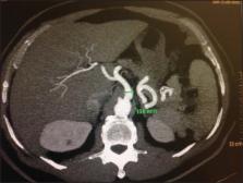
          To analyze the prevalence of anatomical variations of celiac arterial trunk (CAT) branches and hepatic arterial system (HAS), as well as the CAT diameter, length and distance to the superior mesenteric artery.

          Materials and Methods

          Retrospective, cross-sectional and predominantly descriptive study based on the analysis of multidetector computed tomography images of 60 patients.

          Results

          The celiac trunk anatomy was normal in 90% of cases. Hepatosplenic trunk was found in 8.3% of patients, and hepatogastric trunk in 1.7%. Variation of the HAS was observed in 21.7% of cases, including anomalous location of the right hepatic artery in 8.3% of cases, and of the left hepatic artery, in 5%. Also, cases of joint relocation of right and left hepatic arteries, and trifurcation of the proper hepatic artery were observed, respectively, in 3 (5%) and 2 (3.3%) patients. Mean length and caliber of the CAT were 2.3 cm and 0.8 cm, respectively. Mean distance between CAT and superior mesenteric artery was 1.2 cm (standard deviation = 4.08). A significant correlation was observed between CAT diameter and length, and CAT diameter and distance to superior mesenteric artery.

          Conclusion

          The pattern of CAT variations and diameter corroborate the majority of the literature data. However, this does not happen in relation to the HAS.

          Translated abstract

          Objetivo

          Analisar a prevalência de variações anatômicas da ramificação do tronco arterial celíaco (TAC) e do sistema arterial hepático (SAH), o diâmetro e comprimento do TAC e sua distância para a artéria mesentérica superior.

          Materiais e Métodos

          Estudo retrospectivo, transversal, predominantemente descritivo, baseado na análise de imagens de tomografia computadorizada de 60 pacientes.

          Resultados

          A anatomia do TAC foi normal em 90% dos casos. Cinco (8,3%) pacientes apresentaram o tronco hepatoesplênico e um (1,7%) apresentou o tronco hepatogástrico. O SAH variou em 21,7% dos casos. Desses, 8,3% foram na localização anômala da artéria hepática direita e 5% da artéria hepática esquerda. Ainda foram encontrados 3 (5%) casos de relocalização conjunta da artéria hepática direita e artéria hepática esquerda e 2 (3,3%) de trifurcação da artéria hepática própria. A média de comprimento e o calibre médio do TAC foram, respectivamente, 2,33 cm e 0,8 cm. A distância média entre o TAC e a artéria mesentérica superior foi 1,2 cm, com desviopadrão de 4,08. Houve correlação significativa entre diâmetro e comprimento do TAC, e diâmetro do TAC e distância deste para a artéria mesentérica superior.

          Conclusão

          O padrão de variação do TAC e seu diâmetro corroboram a maioria dos dados da literatura, embora o mesmo não tenha ocorrido em relação ao SAH.

          Related collections

          Most cited references28

          • Record: found
          • Abstract: found
          • Article: not found

          Celiac axis and common hepatic artery variations in 5002 patients: systematic analysis with spiral CT and DSA.

          To identify and evaluate the spectrum and prevalence of celiac axis (CA) and common hepatic artery (CHA) variations by using spiral computed tomography (CT) and digital subtraction angiography (DSA). Institutional review board approval was obtained, and the requirement for informed patient consent was waived. The findings in 5002 patients who underwent spiral CT and DSA were retrospectively evaluated. CHA was defined as an arterial trunk containing at least one segmental hepatic artery and the gastroduodenal artery. The pattern of the aortic origin of the branches of the CA and superior mesenteric arteries was analyzed. The CHA anatomy was then investigated. Of 15 possible types of CA variation, 13 types were identified. A normal CA was noted in 4457 (89.1%) of the 5002 patients. Twelve types of CA variation were identified in 482 (9.64%) patients. In the remaining 63 (1.26%) patients, the CA anatomy was classified as ambiguous because the CHA was absent owing to separate origins of the hepatic arteries and the gastroduodenal artery (n = 55) or because the origin of the CHA could not be determined owing to persistent anastomotic channels (n = 8). Seven CHAs originating from the normal CA had a retroportal (n = 6) or transpancreatic (n = 1) course. All eight CHAs originating from the left gastric artery passed the fissure of the ligamentum venosum. The 148 CHAs originating from the superior mesenteric artery showed diverse relationships with the pancreas--being supra-, trans-, or infrapancreatic--and the superior mesenteric-portal venous axis--being pre- or retroportal. The 20 CHAs originating from the aorta had a normal suprapancreatic preportal course. Known or newly found CA and CHA variations could be systematically described in detail. The authors propose a hypothetical anatomic model for summarizing the observed CHA variations. RSNA, 2010
            Bookmark
            • Record: found
            • Abstract: found
            • Article: not found

            Anatomic variations of the hepatic arteries in 604 selective celiac and superior mesenteric angiographies.

            In modern surgical and transplantation procedures the recognition of anatomic vascular abnormalities of the hepatic arteries is of greater importance than ever. The purpose of this study was to evaluate and classify these variations with respect to their impact on visceral surgery. A total of 604 selective celiac and superior mesenteric angiographies performed on patients with known or suspected liver cirrhosis or hepatic or pancreatic malignancies and on donors of partial liver grafts were analyzed retrospectively. The vascular anatomy of the liver was classified according to different established systems and with particular attention to rare variations. Hepatic arterial anatomy as considered normal in textbook descriptions was found in 79.1%, an aberrant or accessory left hepatic artery (LHA) arising from the left gastric artery in 3.0% and an aberrant or accessory right hepatic artery (RHA) branching off the superior mesenteric artery in 11.9% of the cases. In 1.4% of the cases there was a combination of anomalies of both the LHA and RHA. Variants of the celiac trunk, double hepatic arteries branching at the celiac trunk or hepatic arteries arising directly from the aorta, occurred in 4.1% of the cases. Further atypical branches of the LHA and RHA were found in 0.5% of the cases. Since the incidence and pattern of different types of hepatic arterial anatomy can require specialized preoperative diagnostic as well as intraoperative strategies, knowledge of these abnormalities and their frequency is of major importance for the surgeon as well as the radiologist.
              Bookmark
              • Record: found
              • Abstract: found
              • Article: not found

              Anatomical variations of hepatic arterial system, coeliac trunk and renal arteries: an analysis with multidetector CT angiography.

              The purpose of our investigation was to determine the anatomical variations in the coeliac trunk-hepatic arterial system and the renal arteries in patients who underwent multidetector CT (MDCT) angiography of the abdominal aorta for various reasons. A total of 100 patients were analysed retrospectively. The coeliac trunk, hepatic arterial system and renal arteries were analysed individually and anatomical variations were recorded. Statistical analysis of the relationship between hepatocoeliac variations and renal artery variations was performed using a chi(2) test. There was a coeliac trunk trifurcation in 89% and bifurcation in 8% of the cases. Coeliac trunk was absent in 1%, a hepatosplenomesenteric trunk was seen in 1% and a splenomesenteric trunk was present in 1%. Hepatic artery variation was present in 48% of patients. Coeliac trunk and/or hepatic arterial variation was present in 23 (39.7%) of the 58 patients with normal renal arteries, and in 27 (64.3%) of the 42 patients with accessory renal arteries. There was a statistically significant correlation between renal artery variations and coeliac trunk-hepatic arterial system variations (p = 0.015). MDCT angiography permits a correct and detailed evaluation of hepatic and renal vascular anatomy. The prevalence of variations in the coeliac trunk and/or hepatic arteries is increased in people with accessory renal arteries. For that reason, when undertaking angiographic examinations directed towards any single organ, the possibility of variations in the vascular structure of other organs should be kept in mind.
                Bookmark

                Author and article information

                Journal
                Radiol Bras
                Radiol Bras
                rb
                Radiologia Brasileira
                Colégio Brasileiro de Radiologia e Diagnóstico por Imagem
                0100-3984
                1678-7099
                Nov-Dec 2015
                Nov-Dec 2015
                : 48
                : 6
                : 358-362
                Affiliations
                [1 ]PhD, Associate Professor II of Medical Radiology, Universidade Federal da Paraíba (UFPB), João Pessoa, PB, Brazil.
                [2 ]Graduate Students of Medicine at Universidade Federal da Paraíba (UFPB), João Pessoa, PB, Brazil.
                [3 ]PhD, Associate Professor IV of Medical Radiology, Universidade Federal da Paraíba (UFPB), João Pessoa, PB, Brazil.
                [4 ]PhD, Associate Professor II of Anatomy, Universidade Federal da Paraíba (UFPB), João Pessoa, PB, Brazil.
                Author notes
                Mailing Address: Dr. Severino Aires Araujo Neto. Avenida Sapé, 1780, ap. 2201, Bairro Manaíra. João Pessoa, PB, Brazil, 58038-382. E-mail: severinoaires@ 123456hotmail.com .
                Article
                10.1590/0100-3984.2014.0100
                4725396
                26811552
                bdbdcdfc-350c-477a-856c-1519c43ae4c8
                © Colégio Brasileiro de Radiologia e Diagnóstico por Imagem

                This is an Open Access article distributed under the terms of the Creative Commons Attribution License, which permits unrestricted use, distribution, and reproduction in any medium, provided the original work is properly cited.

                History
                : 29 September 2014
                : 31 March 2015
                Categories
                Original Articles

                anatomy,celiac trunk,hepatic artery,multidetector computed tomography

                Comments

                Comment on this article