12
views
0
recommends
+1 Recommend
0 collections
    0
    shares
      • Record: found
      • Abstract: found
      • Article: found
      Is Open Access

      The Role of Ceramides in Insulin Resistance

      review-article

      Read this article at

      Bookmark
          There is no author summary for this article yet. Authors can add summaries to their articles on ScienceOpen to make them more accessible to a non-specialist audience.

          Abstract

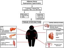
          Resistance to insulin is a pathophysiological state related to the decreased response of peripheral tissues to the insulin action, hyperinsulinemia and raised blood glucose levels caused by increased hepatic glucose outflow. All the above precede the onset of full-blown type 2 diabetes. According to the World Health Organization (WHO), in 2016 more than 1.9 billion people over 18 years of age were overweight and about 600 million were obese. Currently, the primary hypothesis explaining the probability of occurrence of insulin resistance assigns a fundamental role of lipids accumulation in adipocytes or nonadipose tissue (muscle, liver) and the locally developing chronic inflammation caused by adipocytes hypertrophy. However, the major molecular pathways are unknown. The sphingolipid ceramide is the main culprit that combines a plethora of nutrients (e.g., saturated fatty acids) and inflammatory cytokines (e.g., TNFα) to the progression of insulin resistance. The accumulation of sphingolipid ceramide in tissues of obese humans, rodents and Western-diet non-human primates is in line with diabetes, hypertension, cardiac failure or atherosclerosis. In hypertrophied adipose tissue, after adipocytes excel their storage capacity, neutral lipids begin to accumulate in nonadipose tissues, inducing organ dysfunction. Furthermore, obesity is closely related to the development of chronic inflammation and the release of cytokines directly from adipocytes or from macrophages that infiltrate adipose tissue. Enzymes taking part in ceramide metabolism are potential therapeutic targets to manipulate sphingolipids content in tissues, either by inhibition of their synthesis or through stimulation of ceramides degradation. In this review, we will evaluate the mechanisms responsible for the development of insulin resistance and possible therapeutic perspectives.

          Related collections

          Most cited references82

          • Record: found
          • Abstract: found
          • Article: not found

          Monocyte chemoattractant protein 1 in obesity and insulin resistance.

          This study identifies monocyte chemoattractant protein 1 (MCP-1) as an insulin-responsive gene. It also shows that insulin induces substantial expression and secretion of MCP-1 both in vitro in insulin-resistant (IR) 3T3-L1 adipocytes and in vivo in IR obese mice (ob/ob). Thus, MCP-1 resembles other previously described genes (e.g., PAI-1 and SREBP-1c) that remain sensitive to insulin in IR states. The hyperinsulinemia that frequently accompanies obesity and insulin resistance may therefore contribute to the altered expression of these and other genes in insulin target tissues. In vivo studies also demonstrate that MCP-1 is overexpressed in obese mice compared with their lean controls, and that white adipose tissue is a major source of MCP-1. The elevated MCP-1 may alter adipocyte function because addition of MCP-1 to differentiated adipocytes in vitro decreases insulin-stimulated glucose uptake and the expression of several adipogenic genes (LpL, adipsin, GLUT-4, aP2, beta3-adrenergic receptor, and peroxisome proliferator-activated receptor gamma). These results suggest that elevated MCP-1 may induce adipocyte dedifferentiation and contribute to pathologies associated with hyperinsulinemia and obesity, including type II diabetes.
            Bookmark
            • Record: found
            • Abstract: found
            • Article: not found

            An overview of sphingolipid metabolism: from synthesis to breakdown.

            Sphingolipids constitute a class of lipids defined by their eighteen carbon amino-alcohol backbones which are synthesized in the ER from nonsphingolipid precursors. Modification of this basic structure is what gives rise to the vast family of sphingolipids that play significant roles in membrane biology and provide many bioactive metabolites that regulate cell function. Despite the diversity of structure and function of sphingolipids, their creation and destruction are governed by common synthetic and catabolic pathways. In this regard, sphingolipid metabolism can be imagined as an array of interconnected networks that diverge from a single common entry point and converge into a single common breakdown pathway. In their simplest forms, sphingosine, phytosphingosine and dihydrosphingosine serve as the backbones upon which further complexity is achieved. For example, phosphorylation of the C1 hydroxyl group yields the final breakdown products and/or the important signaling molecules sphingosine-1-phosphate, phytosphingosine-1-phosphate and dihydrosphingosine-1-phosphate, respectively. On the other hand, acylation of sphingosine, phytosphingosine, or dihydrosphingosine with one of several possible acyl CoA molecules through the action of distinct ceramide synthases produces the molecules defined as ceramide, phytoceramide, or dihydroceramide. Ceramide, due to the differing acyl CoAs that can be used to produce it, is technically a class of molecules rather than a single molecule and therefore may have different biological functions depending on the acyl chain it is composed of. At the apex of complexity is the group of lipids known as glycosphingolipids (GSL) which contain dozens of different sphingolipid species differing by both the order and type of sugar residues attached to their headgroups. Since these molecules are produced from ceramide precursors, they too may have differences in their acyl chain composition, revealing an additional layer of variation. The glycosphingolipids are divided broadly into two categories: glucosphingolipids and galactosphingolipids. The glucosphingolipids depend initially on the enzyme glucosylceramide synthase (GCS) which attaches glucose as the first residue to the C1 hydroxyl position. Galactosphingolipids, on the other hand, are generated from galactosylceramide synthase (GalCerS), an evolutionarily dissimilar enzyme from GCS. Glycosphingolipids are further divided based upon further modification by various glycosyltransferases which increases the potential variation in lipid species by several fold. Far more abundant are the sphingomyelin species which are produced in parallel with glycosphingolipids, however they are defined by a phosphocholine headgroup rather than the addition of sugar residues. Although sphingomyelin species all share a common headgroup, they too are produced from a variety of ceramide species and therefore can have differing acyl chains attached to their C-2 amino groups. Whether or not the differing acyl chain lengths in SMs dictate unique functions or important biophysical distinctions has not yet been established. Understanding the function of all the existing glycosphingolipids and sphingomyelin species will be a major undertaking in the future since the tools to study and measure these species are only beginning to be developed (see Fig 1 for an illustrated depiction of the various sphingolipid structures). The simple sphingolipids serve both as the precursors and the breakdown products of the more complex ones. Importantly, in recent decades, these simple sphingolipids have gained attention for having significant signaling and regulatory roles within cells. In addition, many tools have emerged to measure the levels of simple sphingolipids and therefore have become the focus of even more intense study in recent years. With this thought in mind, this chapter will pay tribute to the complex sphingolipids, but focus on the regulation of simple sphingolipid metabolism.
              Bookmark
              • Record: found
              • Abstract: found
              • Article: not found
              Is Open Access

              Insulin-stimulated phosphorylation of a Rab GTPase-activating protein regulates GLUT4 translocation.

              Insulin stimulates the rapid translocation of intracellular glucose transporters of the GLUT4 isotype to the plasma membrane in fat and muscle cells. The connections between known insulin signaling pathways and the protein machinery of this membrane-trafficking process have not been fully defined. Recently, we identified a 160-kDa protein in adipocytes, designated AS160, that is phosphorylated by the insulin-activated kinase Akt. This protein contains a GTPase-activating domain (GAP) for Rabs, which are small G proteins required for membrane trafficking. In the present study we have identified six sites of in vivo phosphorylation on AS160. These sites lie in the motif characteristic of Akt phosphorylation, and insulin treatment increased phosphorylation at five of the sites. Expression of AS160 with two or more of these sites mutated to alanine markedly inhibited insulin-stimulated GLUT4 translocation in 3T3-L1 adipocytes. Moreover, this inhibition did not occur when the GAP function in the phosphorylation site mutant was inactivated by a point mutation. These findings strongly indicate that insulin-stimulated phosphorylation of AS160 is required for GLUT4 translocation and that this phosphorylation signals translocation through inactivation of the Rab GAP function.
                Bookmark

                Author and article information

                Contributors
                Journal
                Front Endocrinol (Lausanne)
                Front Endocrinol (Lausanne)
                Front. Endocrinol.
                Frontiers in Endocrinology
                Frontiers Media S.A.
                1664-2392
                21 August 2019
                2019
                : 10
                : 577
                Affiliations
                Department of Hygiene, Epidemiology and Metabolic Disorders, Medical University of Bialystok , Bialystok, Poland
                Author notes

                Edited by: Mohamed Abu-Farha, Dasman Diabetes Institute, Kuwait

                Reviewed by: Asimina Mitrakou-Fanariotou, National and Kapodistrian University of Athens, Greece; Rade Vukovic, The Institute for Health Protection of Mother and Child Serbia, Serbia

                *Correspondence: Emilia Sokolowska emiliasokolowska.umwb@ 123456gmail.com

                This article was submitted to Diabetes, a section of the journal Frontiers in Endocrinology

                Article
                10.3389/fendo.2019.00577
                6712072
                31496996
                bd9f6e5d-c97c-4207-84b4-1ec803cc25ed
                Copyright © 2019 Sokolowska and Blachnio-Zabielska.

                This is an open-access article distributed under the terms of the Creative Commons Attribution License (CC BY). The use, distribution or reproduction in other forums is permitted, provided the original author(s) and the copyright owner(s) are credited and that the original publication in this journal is cited, in accordance with accepted academic practice. No use, distribution or reproduction is permitted which does not comply with these terms.

                History
                : 15 November 2018
                : 07 August 2019
                Page count
                Figures: 6, Tables: 1, Equations: 0, References: 122, Pages: 13, Words: 9796
                Funding
                Funded by: Fundacja na rzecz Nauki Polskiej 10.13039/501100001870
                Funded by: Uniwersytet Medyczny w Bialymstoku 10.13039/501100005297
                Categories
                Endocrinology
                Review

                Endocrinology & Diabetes
                insulin resistance,obesity,ceramide,inflammation,diabetes,therapy
                Endocrinology & Diabetes
                insulin resistance, obesity, ceramide, inflammation, diabetes, therapy

                Comments

                Comment on this article