11
views
0
recommends
+1 Recommend
0 collections
    0
    shares
      • Record: found
      • Abstract: found
      • Article: found
      Is Open Access

      Thrombospondin-4 controls matrix assembly during development and repair of myotendinous junctions

      research-article

      Read this article at

      Bookmark
          There is no author summary for this article yet. Authors can add summaries to their articles on ScienceOpen to make them more accessible to a non-specialist audience.

          Abstract

          Tendons are extracellular matrix (ECM)-rich structures that mediate muscle attachments with the skeleton, but surprisingly little is known about molecular mechanisms of attachment. Individual myofibers and tenocytes in Drosophila interact through integrin (Itg) ligands such as Thrombospondin (Tsp), while vertebrate muscles attach to complex ECM fibrils embedded with tenocytes. We show for the first time that a vertebrate thrombospondin, Tsp4b, is essential for muscle attachment and ECM assembly at myotendinous junctions (MTJs). Tsp4b depletion in zebrafish causes muscle detachment upon contraction due to defects in laminin localization and reduced Itg signaling at MTJs. Mutation of its oligomerization domain renders Tsp4b unable to rescue these defects, demonstrating that pentamerization is required for ECM assembly. Furthermore, injected human TSP4 localizes to zebrafish MTJs and rescues muscle detachment and ECM assembly in Tsp4b-deficient embryos. Thus Tsp4 functions as an ECM scaffold at MTJs, with potential therapeutic uses in tendon strengthening and repair.

          DOI: http://dx.doi.org/10.7554/eLife.02372.001

          eLife digest

          Tendons, the tough connective tissues that link muscles to bones, are essential for lifting, running and other movements in animals. A matrix of proteins, called the extracellular matrix, connects the cells in a tendon, giving it the strength it needs to prevent muscles from detaching from bones during strenuous activities.

          To achieve this strength, extracellular matrix proteins bind to one another and to receptors on the muscle cell surface that are linked to its internal scaffolding, thereby organizing other proteins into a structure called a myotendinous junction. However, despite the essential roles of tendons, scientists do not fully understand how this organization occurs, or how it can go awry.

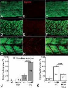
          Subramanian and Schilling screened zebrafish for genes that are essential for proper muscle attachment, and zeroed in on a gene encoding a protein called Thrombospondin-4b (Tsp4b). A similar protein helps to connect muscle and tendon cells in fruit flies. Without Tsp4b, zebrafish are able to form connections between muscles and tendons, but the muscles detach easily during movement. This weakened connection is caused by disorganization of the proteins in the extracellular matrix, which results in reduced signaling from the muscle cell receptors.

          When a human form of this protein was injected into zebrafish embryos lacking Tsp4b, it settled into the junctions between muscle and tendon cells. The human protein repaired the detached muscles and restored the proper organization of the matrix. This improved the strength of the muscle-tendon attachment in the treated fish embryos, suggesting that similar injections could also help to strengthen and repair muscles and tendons in people.

          DOI: http://dx.doi.org/10.7554/eLife.02372.002

          Related collections

          Most cited references76

          • Record: found
          • Abstract: found
          • Article: not found

          RGD and other recognition sequences for integrins.

          Proteins that contain the Arg-Gly-Asp (RGD) attachment site, together with the integrins that serve as receptors for them, constitute a major recognition system for cell adhesion. The RGD sequence is the cell attachment site of a large number of adhesive extracellular matrix, blood, and cell surface proteins, and nearly half of the over 20 known integrins recognize this sequence in their adhesion protein ligands. Some other integrins bind to related sequences in their ligands. The integrin-binding activity of adhesion proteins can be reproduced by short synthetic peptides containing the RGD sequence. Such peptides promote cell adhesion when insolubilized onto a surface, and inhibit it when presented to cells in solution. Reagents that bind selectively to only one or a few of the RGD-directed integrins can be designed by cyclizing peptides with selected sequences around the RGD and by synthesizing RGD mimics. As the integrin-mediated cell attachment influences and regulates cell migration, growth, differentiation, and apoptosis, the RGD peptides and mimics can be used to probe integrin functions in various biological systems. Drug design based on the RGD structure may provide new treatments for diseases such as thrombosis, osteoporosis, and cancer.
            Bookmark
            • Record: found
            • Abstract: found
            • Article: not found

            Thrombospondins are astrocyte-secreted proteins that promote CNS synaptogenesis.

            The establishment of neural circuitry requires vast numbers of synapses to be generated during a specific window of brain development, but it is not known why the developing mammalian brain has a much greater capacity to generate new synapses than the adult brain. Here we report that immature but not mature astrocytes express thrombospondins (TSPs)-1 and -2 and that these TSPs promote CNS synaptogenesis in vitro and in vivo. TSPs induce ultrastructurally normal synapses that are presynaptically active but postsynaptically silent and work in concert with other, as yet unidentified, astrocyte-derived signals to produce functional synapses. These studies identify TSPs as CNS synaptogenic proteins, provide evidence that astrocytes are important contributors to synaptogenesis within the developing CNS, and suggest that TSP-1 and -2 act as a permissive switch that times CNS synaptogenesis by enabling neuronal molecules to assemble into synapses within a specific window of CNS development.
              Bookmark
              • Record: found
              • Abstract: found
              • Article: not found

              Structure of the tendon connective tissue.

              P Kannus (2000)
              Tendons consist of collagen (mostly type I collagen) and elastin embedded in a proteoglycan-water matrix with collagen accounting for 65-80% and elastin approximately 1-2% of the dry mass of the tendon. These elements are produced by tenoblasts and tenocytes, which are the elongated fibroblasts and fibrocytes that lie between the collagen fibers, and are organized in a complex hierarchical scheme to form the tendon proper. Soluble tropocollagen molecules form cross-links to create insoluble collagen molecules which then aggregate progressively into microfibrils and then into electronmicroscopically clearly visible units, the collagen fibrils. A bunch of collagen fibrils forms a collagen fiber, which is the basic unit of a tendon. A fine sheath of connective tissue called endotenon invests each collagen fiber and binds fibers together. A bunch of collagen fibers forms a primary fiber bundle, and a group of primary fiber bundles forms a secondary fiber bundle. A group of secondary fiber bundles, in turn, forms a tertiary bundle, and the tertiary bundles make up the tendon. The entire tendon is surrounded by a fine connective tissue sheath called epitenon. The three-dimensional ultrastructure of tendon fibers and fiber bundles is complex. Within one collagen fiber, the fibrils are oriented not only longitudinally but also transversely and horizontally. The longitudinal fibers do not run only parallel but also cross each other, forming spirals. Some of the individual fibrils and fibril groups form spiral-type plaits. The basic function of the tendon is to transmit the force created by the muscle to the bone, and, in this way, make joint movement possible. The complex macro- and microstructure of tendons and tendon fibers make this possible. During various phases of movements, the tendons are exposed not only to longitudinal but also to transversal and rotational forces. In addition, they must be prepared to withstand direct contusions and pressures. The above-described three-dimensional internal structure of the fibers forms a buffer medium against forces of various directions, thus preventing damage and disconnection of the fibers.
                Bookmark

                Author and article information

                Contributors
                URI : http://orcid.org/0000-0001-8455-6804
                Role: Reviewing editor
                Journal
                eLife
                Elife
                eLife
                eLife
                eLife
                eLife Sciences Publications, Ltd
                2050-084X
                18 June 2014
                2014
                : 3
                : e02372
                Affiliations
                [1 ]Department of Developmental and Cell Biology, University of California, Irvine , Irvine, United States
                University of Sheffield , United Kingdom
                University of Sheffield , United Kingdom
                Author notes
                [* ]For correspondence: tschilli@ 123456uci.edu
                Article
                02372
                10.7554/eLife.02372
                4096842
                24941943
                bd4534e0-0431-4e23-b570-99398ac1df45
                Copyright © 2014, Subramanian and Schilling

                This article is distributed under the terms of the Creative Commons Attribution License, which permits unrestricted use and redistribution provided that the original author and source are credited.

                History
                : 22 January 2014
                : 17 June 2014
                Funding
                Funded by: National Institutes of Health (NIH) FundRef identification ID: http://dx.doi.org/10.13039/100000002
                Award ID: R21 AR62792, R01 DE13828
                Award Recipient :
                The funder had no role in study design, data collection and interpretation, or the decision to submit the work for publication.
                Categories
                Research Article
                Cell Biology
                Developmental Biology and Stem Cells
                Custom metadata
                0.7
                Functional attachments between muscles and tendons require pentameric Thrombospondin-4, revealing novel roles both as an integrin ligand and extracellular matrix scaffold.

                Life sciences
                myotendinous junctions,muscle attachment,ecm organization,tendon development,zebrafish

                Comments

                Comment on this article