3
views
0
recommends
+1 Recommend
0 collections
    0
    shares
      • Record: found
      • Abstract: found
      • Article: found
      Is Open Access

      Inflammatory myopathy with myasthenia gravis : Thymoma association and polymyositis pathology

      research-article

      Read this article at

      Bookmark
          There is no author summary for this article yet. Authors can add summaries to their articles on ScienceOpen to make them more accessible to a non-specialist audience.

          Abstract

          Objective

          To provide evidence that idiopathic inflammatory myopathy (IM) with myasthenia gravis (MG) frequently shows thymoma association and polymyositis (PM) pathology and shares clinicopathologic characteristics with IM induced by immune checkpoint inhibitors (ICIs).

          Methods

          We analyzed the clinicopathologic features of 10 patients with idiopathic IM and MG identified in 970 consecutive patients with biopsy-proven IM.

          Results

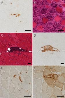
          Seven patients (70%) had thymoma. IM and MG were diagnosed with more than 5-year time difference in 6 thymomatous patients and within 1 year in 1 thymomatous and 3 nonthymomatous patients. Seven thymomatous patients showed rhabdomyolysis-like features with respiratory failure (4/7), dropped head (3/7), cardiac involvement (2/7), and positive anti–acetylcholine receptor (anti-AChR) and anti-titin antibodies (7/7 and 4/6, respectively) but rarely showed ocular symptoms (2/7) or decremental repetitive nerve stimulation (RNS) responses (1/7) at IM diagnosis. Three nonthymomatous patients showed acute cardiorespiratory failure with rhabdomyolysis-like features (1/3), positive anti-AChR and anti-titin antibodies (3/2 and 2/2, respectively), and fluctuating weakness of the skeletal muscle without ocular symptoms (3/3). Muscle pathology showed a PM pathology with infiltration of CD8-positive CD45RA-negative T-lymphocytes (9/9), scattered endomysial programmed cell death 1 (PD-1)–positive cells (9/9), and overexpression of programmed cell death ligand 1 (PD-L1) on the sarcolemma of muscle fibers around the infiltrating PD-1–positive cells (7/9).

          Conclusion

          Rhabdomyolysis-like features, positive anti-AChR antibody without decremental RNS responses, and PD-L1 overexpression are possible characteristics shared by ICI-induced IM. Frequent thymoma association in patients with idiopathic IM and MG may suggest thymoma-related immunopathogenic mechanisms, including dysregulation of the immune checkpoint pathway.

          Related collections

          Most cited references28

          • Record: found
          • Abstract: found
          • Article: found
          Is Open Access

          CTLA-4 and PD-1 Pathways

          Supplemental Digital Content is available in the text.
            Bookmark
            • Record: found
            • Abstract: found
            • Article: not found

            Atypical neurological complications of ipilimumab therapy in patients with metastatic melanoma.

            Ipilimumab is a novel FDA-approved recombinant human monoclonal antibody that blocks cytotoxic T-lymphocyte antigen-4 and has been used to treat patients with metastatic melanoma. Immune-related neurological adverse effects include inflammatory myopathy, aseptic meningitis, posterior reversible encephalopathy syndrome, Guillain-Barré syndrome, myasthenia gravis-type syndrome, sensorimotor neuropathy, and inflammatory enteric neuropathy. To date, there is no report for ipilimumab-induced chronic inflammatory demyelinating polyneuropathy (CIDP), transverse myelitis (TM), or concurrent myositis and myasthenia gravis-type syndrome. Our objective is to raise early recognition of atypical neurological adverse events and to share our therapeutic approach.
              Bookmark
              • Record: found
              • Abstract: found
              • Article: found
              Is Open Access

              Myasthenic crisis and polymyositis induced by one dose of nivolumab

              An 80‐year‐old man, who developed multiple lymph node and skin metastasis of malignant melanoma, received nivolumab monotherapy. Two weeks after the first dose, he experienced anorexia and fatigue, and suffered from progressive, severe dyspnea and muscle weakness. We diagnosed him with myocarditis, myositis, and myasthenic crisis induced by nivolumab. We commenced steroid therapy, immune absorption therapy, plasma exchange therapy, and i.v. immunoglobulin therapy, and succeeded in saving his life. Because his serum level of anti‐acetylcholine receptor antibodies in a sample collected before nivolumab treatment were positive and were elevated significantly after nivolumab, we suspected that nivolumab triggered a severe autoimmune response, which progressed subclinical myasthenia gravis to myasthenic crisis. We carried out T cell receptor repertoire analysis using next‐generation sequencing technologies and identified infiltration of clonally expanded T cell populations in the skeletal muscle after nivolumab treatment, implying a very strong T cell immune response against muscular cells. To avoid severe immune‐related adverse events, the exclusion of patients with subclinical autoimmune disease is very important for treatment with immune checkpoint inhibitors.
                Bookmark

                Author and article information

                Contributors
                Journal
                Neurol Neuroimmunol Neuroinflamm
                Neurol Neuroimmunol Neuroinflamm
                nnn
                NEURIMMINFL
                Neurology® Neuroimmunology & Neuroinflammation
                Lippincott Williams & Wilkins (Hagerstown, MD )
                2332-7812
                24 December 2018
                March 2019
                24 December 2018
                : 6
                : 2
                : e535
                Affiliations
                From the Department of Neurology (N.U., K.T., C.I., A.U., A.K., T.T., J.S.), Graduate School of Medicine, University of Tokyo; Division of Neurology (M.K., K.K.), Department of Internal Medicine, National Defense Medical College, Saitama; Division of Neurology (Y. Shiio), Tokyo Teishin Hospital; Department of Neurology (Y. Sakurai), Mitsui Memorial Hospital; Department of Neurology (Y.H.), Teikyo University School of Medicine; and Department of Neurology (K.Y., S.N.-E.), Fukushima Medical University, Japan.
                Author notes
                Correspondence Dr. Shimizu jshimizu-tky@ 123456umin.ac.jp

                Funding information and disclosures are provided at the end of the article. Full disclosure form information provided by the authors is available with the full text of this article at Neurology.org/NN.

                The Article Processing Charge was funded by the authors.

                Article
                NEURIMMINFL2018018416
                10.1212/NXI.0000000000000535
                6340335
                30697585
                bcedd898-db61-4cb5-8ae9-b2d5a1a365e1
                Copyright © 2018 The Author(s). Published by Wolters Kluwer Health, Inc. on behalf of the American Academy of Neurology.

                This is an open access article distributed under the terms of the Creative Commons Attribution-NonCommercial-NoDerivatives License 4.0 (CC BY-NC-ND), which permits downloading and sharing the work provided it is properly cited. The work cannot be changed in any way or used commercially without permission from the journal.

                History
                : 04 September 2018
                : 24 October 2018
                Categories
                132
                185
                Article
                Custom metadata
                TRUE

                Comments

                Comment on this article