0
views
0
recommends
+1 Recommend
0 collections
    0
    shares
      • Record: found
      • Abstract: found
      • Article: found
      Is Open Access

      Differences in artificial ligament graft osseointegration of the anterior cruciate ligament in a sheep model: a comparison between interference screw and cortical suspensory fixation

      research-article

      Read this article at

      Bookmark
          There is no author summary for this article yet. Authors can add summaries to their articles on ScienceOpen to make them more accessible to a non-specialist audience.

          Abstract

          Background

          Interference screws are the most common femoral fixation for anterior cruciate ligament (ACL) reconstruction with polyethylene terephthalate (PET) artificial ligaments. However, interference screws have several disadvantages, such as the risk of one tunnel blowout and damage to the graft. Suspensory fixations have the advantages of high tensile strength and promotion of graft bone contact. The purpose of this study was to compare PET artificial ligament graft osseointegration between interference screw fixation (ISF) and cortical suspensory fixation (CSF) for ACL reconstruction.

          Methods

          Forty sheep underwent ACL reconstruction of the right knee with PET artificial ligament. The graft was fixed with ISF or CSF for femoral fixation. Animals were randomly assigned to the ISF (n=20) or the CSF (n=20) groups. The sheep were sacrificed at 3 or 12 months postoperatively for biomechanical tests, micro computed tomography (micro-CT) scans, and histological assessments.

          Results

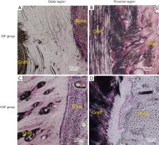
          The mean load-to-failure between the CSF group (836±355 N) appeared higher than that of the ISF group (604±277 N) at 3 months, but no significant difference was detected between the groups (P=0.24). At 12 months, there was also no significant difference in load-to-failure between the CSF and ISF groups (1,194±350 vs. 1,097±764 N; P=0.78). According to the micro-CT scan results, the femoral bone tunnel diameter of the ISF group appeared larger than that of the CSF group at 3 months (12±1 vs. 10±1 mm; P=0.02) and similar to that of the CSF group at 12 months (12±1 vs. 11±2 mm; P=0.38). Furthermore, histological results showed that at the graft-tunnel interface of the femoral tunnel aperture, disoriented fibers formed in the ISF group while oriented and dense fibers formed in the CSF group.

          Conclusions

          ACLR with synthetic ligament by cortical suspension devices with adjustable loops demonstrated a better graft-bone healing capacity at the femoral tunnel aperture compared with that from titanium interference screws over 12 months postoperatively. No significant difference was found in biomechanical strength between the two fixation methods during the early healing stage.

          Related collections

          Most cited references45

          • Record: found
          • Abstract: found
          • Article: not found

          Donor-site morbidity and anterior knee problems after anterior cruciate ligament reconstruction using autografts.

          The authors review the current knowledge on donor site-related problems after using different types of autografts for anterior cruciate ligament (ACL) reconstruction and make recommendations on minimizing late donor-site problems. Postoperative donor-site morbidity and anterior knee pain following ACL surgery may result in substantial impairment for patients. The selection of graft, surgical technique, and rehabilitation program can affect the severity of pain that patients experience. The loss or disturbance of anterior sensitivity caused by intraoperative injury to the infrapatellar nerve(s) in conjunction with patellar tendon harvest is correlated with donor-site discomfort and an inability to kneel and knee-walk. The patellar tendon at the donor site has significant clinical, radiographic, and histologic abnormalities 2 years after harvest of its central third. Donor-site discomfort correlates poorly with radiographic and histologic findings after the use of patellar tendon autografts. The use of hamstring tendon autografts appears to cause less postoperative donor-site morbidity and anterior knee problems than the use of patellar tendon autografts. There also appears to be a regrowth of the hamstring tendons within 2 years of the harvesting procedure. There is little known about the effect on the donor site of harvesting fascia lata and quadriceps tendon autografts. Efforts should be made to spare the infrapatellar nerve(s) during ACL reconstruction using patellar tendon autografts. Reharvesting the patellar tendon cannot be recommended due to significant clinical, radiographic, and histologic abnormalities 2 years after harvesting its central third. It is important to regain full range of motion and strength after the use of any type of autograft to avoid future anterior knee problems. If randomized controlled trials show that the long-term laxity measurements following ACL reconstruction using hamstring tendon autografts are equal to those of patellar tendon autografts, we recommend the use of hamstring tendon autografts because there are fewer donor-site problems.
            Bookmark
            • Record: found
            • Abstract: found
            • Article: not found

            Bone tunnel enlargement after anterior cruciate ligament reconstruction: fact or fiction?

            Radiographic enlargement of bone tunnels following anterior cruciate ligament (ACL) reconstruction has been recently introduced in the literature; however, the etiology and clinical relevance of this phenomenon remain unclear. While early reports suggested that bone tunnel enlargement is mainly the result of an immune response to allograft tissue, more recent studies imply that other biological as well as mechanical factors play a more important role. Biological factors associated with tunnel enlargement include foreign-body immune response (against allografts), non-specific inflammatory response (as in osteolysis around total joint implants), cell necrosis due to toxic products in the tunnel (ethylene oxide, metal), and heat necrosis as a response to drilling (natural course). Mechanical factors contributing to tunnel enlargement include stress deprivation of bone within the tunnel wall, graft-tunnel motion, improper tunnel placement, and aggressive rehabilitation. Graft-tunnel motion refers to longitudinal and transverse motion of the graft within the bone tunnel and can occur with various graft types and fixation techniques. Aggressive rehabilitation programmes may contribute to tunnel enlargement as the graft-bone interface is subjected to early stress before biological incorporation is complete. Further basic research is required to verify the effect of the various proposed factors on the etiology of bone tunnel enlargement. We recommend that routine follow-up examinations after ACL reconstruction should include the measurement of bone tunnel size in order to contribute to a better understanding of the incidence, time course, and clinical relevance of this phenomenon. Improved and more anatomical surgical fixation techniques may be useful for the prevention of bone tunnel enlargement.
              Bookmark
              • Record: found
              • Abstract: found
              • Article: not found

              Tendon healing in a bone tunnel differs at the tunnel entrance versus the tunnel exit: an effect of graft-tunnel motion?

              Motion between a tendon graft and bone tunnel may impair graft incorporation and lead to tunnel widening. Healing of a tendon graft in a bone tunnel is inhibited by graft-tunnel motion. Controlled laboratory study. Anterior cruciate ligament reconstruction was performed in 5 cadaveric rabbit limbs, and 3-dimensional graft-tunnel motion was measured using micro-computed tomography. The authors then performed bilateral anterior cruciate ligament reconstruction in 15 rabbits and used histomorphometry to compare tendon-to-bone healing between the tunnel aperture, midtunnel, and tunnel exit and between the anterior and posterior aspects of the tunnel. Graft-tunnel motion was greatest at the tunnel apertures and least at the tunnel exit in cadaveric testing. Healing of the graft was slowest at the tunnel apertures. Tendon-bone interface width was greater at the aperture than at the tunnel exit for the femoral tunnel (P = .04). There was an inverse correlation between time zero graft-tunnel motion and healing in the femoral tunnel (P = .005). There was closer apposition of new bone to the tendon graft in the posterior half of the interface (P < .05). Osteoclasts were found at the tunnel apertures. Although graft-tunnel motion was only measured in cadaveric animals, results suggest that healing may be affected by the local mechanical environment, as graft healing in the femoral tunnel was inversely proportional to the magnitude of graft-tunnel motion. Graft-tunnel motion may impair early graft incorporation and may lead to osteoclast-mediated bone resorption, contributing to tunnel widening. Early, aggressive postoperative rehabilitation may have detrimental effects on graft-to-bone healing.
                Bookmark

                Author and article information

                Journal
                Ann Transl Med
                Ann Transl Med
                ATM
                Annals of Translational Medicine
                AME Publishing Company
                2305-5839
                2305-5847
                September 2021
                September 2021
                : 9
                : 17
                : 1370
                Affiliations
                [1]deptDepartment of Sports Medicine , Huashan Hospital , Shanghai, China
                Author notes

                Contributions: (I) Conception and design: H Li, F Jiang, Y Ge, S Chen; (II) Administrative support: S Chen; (III) Provision of study materials or patients: H Li, H Li, F Wan; (IV) Collection and assembly of data: H Li, F Jiang, H Li, Y Ge; (V) Data analysis and interpretation: H Li, F Jiang, Y Ge; (VI) Manuscript writing: All authors; (VII) Final approval of manuscript: All authors.

                [#]

                These authors contributed equally to this work.

                Correspondence to: Shiyi Chen, MD, PhD. No. 12, Wulumuqi Zhong Road, Shanghai 200040, China. Email: cshiyi@ 123456163.com .
                Article
                atm-09-17-1370
                10.21037/atm-21-1076
                8506542
                34733922
                bcb690fa-95be-4ced-8d1c-357bad921d42
                2021 Annals of Translational Medicine. All rights reserved.

                Open Access Statement: This is an Open Access article distributed in accordance with the Creative Commons Attribution-NonCommercial-NoDerivs 4.0 International License (CC BY-NC-ND 4.0), which permits the non-commercial replication and distribution of the article with the strict proviso that no changes or edits are made and the original work is properly cited (including links to both the formal publication through the relevant DOI and the license). See: https://creativecommons.org/licenses/by-nc-nd/4.0.

                History
                : 07 March 2021
                : 14 July 2021
                Categories
                Original Article

                anterior cruciate ligament (acl),artificial ligament,cortical suspensory fixation (csf),interference screw fixation (isf),graft osseointegration

                Comments

                Comment on this article