8
views
0
recommends
+1 Recommend
0 collections
    0
    shares
      • Record: found
      • Abstract: found
      • Article: found
      Is Open Access

      Laminin γ3 plays an important role in retinal lamination, photoreceptor organisation and ganglion cell differentiation

      research-article

      Read this article at

      Bookmark
          There is no author summary for this article yet. Authors can add summaries to their articles on ScienceOpen to make them more accessible to a non-specialist audience.

          Abstract

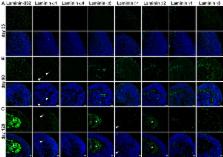
          Laminins are heterotrimeric glycoproteins of the extracellular matrix. Eleven different laminin chains have been identified in vertebrates. They are ubiquitously expressed in the human body, with a distinct tissue distribution. Laminin expression in neural retina and their functional role during human retinogenesis is still unknown. This study investigated the laminin expression in human developing and adult retina, showing laminin α1, α5, β1, β2 and γ1 to be predominantly expressed in Bruch’s membrane and the inner limiting membrane. Laminin-332 and laminin γ3 expression were mainly observed in the neural retina during retinal histogenesis. These expression patterns were largely conserved in pluripotent stem cell-derived retinal organoids. Blocking of laminin γ3 function in retinal organoids resulted in the disruption of laminar organisation and synapse formation, the loss of photoreceptor organisation and retinal ganglion cells. Our data demonstrate a unique temporal and spatial expression for laminins and reveal a novel role for laminin γ3 during human retinogenesis.

          Related collections

          Most cited references46

          • Record: found
          • Abstract: found
          • Article: not found

          A simplified laminin nomenclature.

          A simplification of the laminin nomenclature is presented. Laminins are multidomain heterotrimers composed of alpha, beta and gamma chains. Previously, laminin trimers were numbered with Arabic numerals in the order discovered, that is laminins-1 to -5. We introduce a new identification system for a trimer using three Arabic numerals, based on the alpha, beta and gamma chain numbers. For example, the laminin with the chain composition alpha5beta1gamma1 is termed laminin-511, and not laminin-10. The current practice is also to mix two overlapping domain and module nomenclatures. Instead of the older Roman numeral nomenclature and mixed nomenclature, all modules are now called domains. Some domains are renamed or renumbered. Laminin epidermal growth factor-like (LE) domains are renumbered starting at the N-termini, to be consistent with general protein nomenclature. Domain IVb of alpha chains is named laminin 4a (L4a), domain IVa of alpha chains is named L4b, domain IV of gamma chains is named L4, and domain IV of beta chains is named laminin four (LF). The two coiled-coil domains I and II are now considered one laminin coiled-coil domain (LCC). The interruption in the coiled-coil of beta chains is named laminin beta-knob (Lbeta) domain. The chain origin of a domain is specified by the chain nomenclature, such as alpha1L4a. The abbreviation LM is suggested for laminin. Otherwise, the nomenclature remains unaltered.
            Bookmark
            • Record: found
            • Abstract: found
            • Article: not found

            Functional diversity of laminins.

            Laminins are a large family of conserved, multidomain trimeric basement membrane proteins that contribute to the structure of extracellular matrix and influence the behavior of associated cells, such as adhesion, differentiation, migration, phenotype stability, and resistance to anoikis. In lower organisms such as Hydra there is only one isoform of laminin, but higher organisms have at least 16 trimeric isoforms with varying degrees of cell/tissue specificity. In vitro protein and cell culture studies, gene manipulation in animals, and laminin gene mutations in human diseases have provided insight into the specific functions of some laminins, but the biological roles of many isoforms are still largely unexplored, mainly owing to difficulties in isolating them in pure form from tissues or cells. In this review, we elucidate the evolution of laminins, describe their molecular complexity, and explore the current knowledge of their diversity and functional aspects, including laminin-mediated signaling via membrane receptors, in vitro cell biology, and involvement in various tissues gained from animal model and human disease studies. The potential use of laminins in cell biology research and biotechnology is discussed.
              Bookmark
              • Record: found
              • Abstract: found
              • Article: not found

              Generation of a ciliary margin-like stem cell niche from self-organizing human retinal tissue.

              In the developing neural retina (NR), multipotent stem cells within the ciliary margin (CM) contribute to de novo retinal tissue growth. We recently reported the ability of human embryonic stem cells (hESCs) to self-organize stratified NR using a three-dimensional culture technique. Here we report the emergence of CM-like stem cell niches within human retinal tissue. First, we developed a culture method for selective NR differentiation by timed BMP4 treatment. We then found that inhibiting GSK3 and FGFR induced the transition from NR tissue to retinal pigment epithelium (RPE), and that removing this inhibition facilitated the reversion of this RPE-like tissue back to the NR fate. This step-wise induction-reversal method generated tissue aggregates with RPE at the margin of central-peripherally polarized NR. We demonstrate that the NR-RPE boundary tissue further self-organizes a niche for CM stem cells that functions to expand the NR peripherally by de novo progenitor generation.
                Bookmark

                Author and article information

                Contributors
                +44-191-241-8688 , majlinda.lako@ncl.ac.uk
                Journal
                Cell Death Dis
                Cell Death Dis
                Cell Death & Disease
                Nature Publishing Group UK (London )
                2041-4889
                23 May 2018
                23 May 2018
                June 2018
                : 9
                : 6
                : 615
                Affiliations
                [1 ]ISNI 0000 0001 0462 7212, GRID grid.1006.7, Institute of Genetic Medicine, , Newcastle University, ; Newcastle upon Tyne, United Kingdom
                [2 ]ISNI 0000 0001 0462 7212, GRID grid.1006.7, Institute of Neuroscience, , Newcastle University, ; Newcastle upon Tyne, United Kingdom
                [3 ]ISNI 0000 0004 1936 7910, GRID grid.1012.2, Lions Eye Institute, Centre for Ophthalmology and Visual Science, , University of Western Australia, ; Nedlands WA, Australia
                Author information
                http://orcid.org/0000-0001-9165-3020
                http://orcid.org/0000-0003-3685-6516
                Article
                648
                10.1038/s41419-018-0648-0
                5966411
                29795281
                bc2f8911-94d0-4e85-b63f-aa27ab26efa6
                © The Author(s) 2018

                Open Access This article is licensed under a Creative Commons Attribution 4.0 International License, which permits use, sharing, adaptation, distribution and reproduction in any medium or format, as long as you give appropriate credit to the original author(s) and the source, provide a link to the Creative Commons license, and indicate if changes were made. The images or other third party material in this article are included in the article’s Creative Commons license, unless indicated otherwise in a credit line to the material. If material is not included in the article’s Creative Commons license and your intended use is not permitted by statutory regulation or exceeds the permitted use, you will need to obtain permission directly from the copyright holder. To view a copy of this license, visit http://creativecommons.org/licenses/by/4.0/.

                History
                : 6 March 2018
                : 12 April 2018
                : 26 April 2018
                Categories
                Article
                Custom metadata
                © The Author(s) 2018

                Cell biology
                Cell biology

                Comments

                Comment on this article