44
views
0
recommends
+1 Recommend
0 collections
    0
    shares
      • Record: found
      • Abstract: found
      • Article: found
      Is Open Access

      Imaging the where and when of tic generation and resting state networks in adult Tourette patients

      research-article

      Read this article at

      Bookmark
          There is no author summary for this article yet. Authors can add summaries to their articles on ScienceOpen to make them more accessible to a non-specialist audience.

          Abstract

          Introduction: Tourette syndrome (TS) is a neuropsychiatric disorder with the core phenomenon of tics, whose origin and temporal pattern are unclear. We investigated the When and Where of tic generation and resting state networks (RSNs) via functional magnetic resonance imaging (fMRI).

          Methods: Tic-related activity and the underlying RSNs in adult TS were studied within one fMRI session. Participants were instructed to lie in the scanner and to let tics occur freely. Tic onset times, as determined by video-observance were used as regressors and added to preceding time-bins of 1 s duration each to detect prior activation. RSN were identified by independent component analysis (ICA) and correlated to disease severity by the means of dual regression.

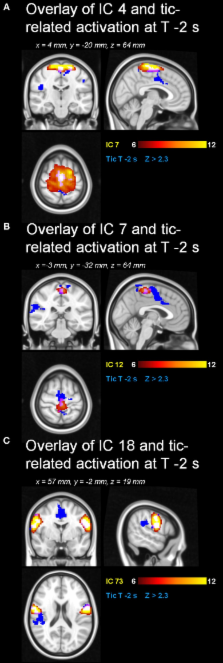
          Results: Two seconds before a tic, the supplementary motor area (SMA), ventral primary motor cortex, primary sensorimotor cortex and parietal operculum exhibited activation; 1 s before a tic, the anterior cingulate, putamen, insula, amygdala, cerebellum and the extrastriatal-visual cortex exhibited activation; with tic-onset, the thalamus, central operculum, primary motor and somatosensory cortices exhibited activation. Analysis of resting state data resulted in 21 components including the so-called default-mode network. Network strength in those regions in SMA of two premotor ICA maps that were also active prior to tic occurrence, correlated significantly with disease severity according to the Yale Global Tic Severity Scale (YGTTS) scores.

          Discussion: We demonstrate that the temporal pattern of tic generation follows the cortico-striato-thalamo-cortical circuit, and that cortical structures precede subcortical activation. The analysis of spontaneous fluctuations highlights the role of cortical premotor structures. Our study corroborates the notion of TS as a network disorder in which abnormal RSN activity might contribute to the generation of tics in SMA.

          Related collections

          Most cited references62

          • Record: found
          • Abstract: found
          • Article: not found

          Greater than the sum of its parts: a review of studies combining structural connectivity and resting-state functional connectivity.

          It is commonly assumed that functional brain connectivity reflects structural brain connectivity. The exact relationship between structure and function, however, might not be straightforward. In this review we aim to examine how our understanding of the relationship between structure and function in the 'resting' brain has advanced over the last several years. We discuss eight articles that directly compare resting-state functional connectivity with structural connectivity and three clinical case studies of patients with limited white matter connections between the cerebral hemispheres. All studies examined show largely convergent results: the strength of resting-state functional connectivity is positively correlated with structural connectivity strength. However, functional connectivity is also observed between regions where there is little or no structural connectivity, which most likely indicates functional correlations mediated by indirect structural connections (i.e. via a third region). As the methodologies for measuring structural and functional connectivity continue to improve and their complementary strengths are applied in parallel, we can expect important advances in our diagnostic and prognostic capacities in diseases like Alzheimer's, multiple sclerosis, and stroke.
            Bookmark
            • Record: found
            • Abstract: found
            • Article: not found

            Reliable intrinsic connectivity networks: test-retest evaluation using ICA and dual regression approach.

            Functional connectivity analyses of resting-state fMRI data are rapidly emerging as highly efficient and powerful tools for in vivo mapping of functional networks in the brain, referred to as intrinsic connectivity networks (ICNs). Despite a burgeoning literature, researchers continue to struggle with the challenge of defining computationally efficient and reliable approaches for identifying and characterizing ICNs. Independent component analysis (ICA) has emerged as a powerful tool for exploring ICNs in both healthy and clinical populations. In particular, temporal concatenation group ICA (TC-GICA) coupled with a back-reconstruction step produces participant-level resting state functional connectivity maps for each group-level component. The present work systematically evaluated the test-retest reliability of TC-GICA derived RSFC measures over the short-term (<45 min) and long-term (5-16 months). Additionally, to investigate the degree to which the components revealed by TC-GICA are detectable via single-session ICA, we investigated the reproducibility of TC-GICA findings. First, we found moderate-to-high short- and long-term test-retest reliability for ICNs derived by combining TC-GICA and dual regression. Exceptions to this finding were limited to physiological- and imaging-related artifacts. Second, our reproducibility analyses revealed notable limitations for template matching procedures to accurately detect TC-GICA based components at the individual scan level. Third, we found that TC-GICA component's reliability and reproducibility ranks are highly consistent. In summary, TC-GICA combined with dual regression is an effective and reliable approach to exploratory analyses of resting state fMRI data. Copyright (c) 2009 Elsevier Inc. All rights reserved.
              Bookmark
              • Record: found
              • Abstract: found
              • Article: not found

              Contributions of anterior cingulate cortex to behaviour.

              Assessments of anterior cingulate cortex in experimental animals and humans have led to unifying theories of its structural organization and contributions to mammalian behaviour. The anterior cingulate cortex forms a large region around the rostrum of the corpus callosum that is termed the anterior executive region. This region has numerous projections into motor systems, however, since these projections originate from different parts of anterior cingulate cortex and because functional studies have shown that it does not have a uniform contribution to brain functions, the anterior executive region is further subdivided into 'affect' and 'cognition' components. The affect division includes areas 25, 33 and rostral area 24, and has extensive connections with the amygdala and periaqueductal grey, and parts of it project to autonomic brainstem motor nuclei. In addition to regulating autonomic and endocrine functions, it is involved in conditioned emotional learning, vocalizations associated with expressing internal states, assessments of motivational content and assigning emotional valence to internal and external stimuli, and maternal-infant interactions. The cognition division includes caudal areas 24' and 32', the cingulate motor areas in the cingulate sulcus and nociceptive cortex. The cingulate motor areas project to the spinal cord and red nucleus and have premotor functions, while the nociceptive area is engaged in both response selection and cognitively demanding information processing. The cingulate epilepsy syndrome provides important support of experimental animal and human functional imaging studies for the role of anterior cingulate cortex in movement, affect and social behaviours. Excessive cingulate activity in cases with seizures confirmed in anterior cingulate cortex with subdural electrode recordings, can impair consciousness, alter affective state and expression, and influence skeletomotor and autonomic activity. Interictally, patients with anterior cingulate cortex epilepsy often display psychopathic or sociopathic behaviours. In other clinical examples of elevated anterior cingulate cortex activity it may contribute to tics, obsessive-compulsive behaviours, and aberrent social behaviour. Conversely, reduced cingulate activity following infarcts or surgery can contribute to behavioural disorders including akinetic mutism, diminished self-awareness and depression, motor neglect and impaired motor initiation, reduced responses to pain, and aberrent social behaviour. The role of anterior cingulate cortex in pain responsiveness is suggested by cingulumotomy results and functional imaging studies during noxious somatic stimulation. The affect division of anterior cingulate cortex modulates autonomic activity and internal emotional responses, while the cognition division is engaged in response selection associated with skeletomotor activity and responses to noxious stimuli. Overall, anterior cingulate cortex appears to play a crucial role in initiation, motivation, and goal-directed behaviours.(ABSTRACT TRUNCATED AT 400 WORDS)
                Bookmark

                Author and article information

                Contributors
                Journal
                Front Hum Neurosci
                Front Hum Neurosci
                Front. Hum. Neurosci.
                Frontiers in Human Neuroscience
                Frontiers Media S.A.
                1662-5161
                28 May 2014
                2014
                : 8
                : 362
                Affiliations
                [1] 1Institute of Neuroscience and Medicine - 4, Forschungszentrum Jülich GmbH Jülich, Germany
                [2] 2Department of Psychiatry, Psychotherapy and Psychosomatics, RWTH Aachen University Aachen, Germany
                [3] 3JARA – Translational Brain Medicine Aachen, Germany
                [4] 4Department of Neurology, RWTH Aachen University Aachen, Germany
                Author notes

                Edited by: John J. Foxe, Albert Einstein College of Medicine, USA

                Reviewed by: Sheng Zhang, Yale University, USA; Rupert Lanzenberger, Medical University of Vienna, Austria; Agatha Lenartowicz, University of California Los Angeles, USA

                *Correspondence: Irene Neuner, Institute of Neuroscience and Medicine – 4, Forschungszentrum Jülich GmbH, Leo-Brandt-Straße, 52425 Jülich, Germany e-mail: i.neuner@ 123456fz-juelich.de

                This article was submitted to the journal Frontiers in Human Neuroscience.

                †Present address: Tony Stöcker, DZNE, Bonn, Germany

                ‡These authors have contributed equally to this work.

                Article
                10.3389/fnhum.2014.00362
                4035756
                24904391
                bb9853e1-64b2-46b2-a74c-2b83a405aaff
                Copyright © 2014 Neuner, Werner, Arrubla, Stöcker, Ehlen, Wegener, Schneider and Shah.

                This is an open-access article distributed under the terms of the Creative Commons Attribution License (CC BY). The use, distribution or reproduction in other forums is permitted, provided the original author(s) or licensor are credited and that the original publication in this journal is cited, in accordance with accepted academic practice. No use, distribution or reproduction is permitted which does not comply with these terms.

                History
                : 08 November 2013
                : 12 May 2014
                Page count
                Figures: 7, Tables: 2, Equations: 0, References: 88, Pages: 16, Words: 11515
                Categories
                Neuroscience
                Original Research Article

                Neurosciences
                resting state networks,tourette,tics,basal ganglia,cortico-striato-thalamo-cortical circuit

                Comments

                Comment on this article