26
views
0
recommends
+1 Recommend
0 collections
    0
    shares
      • Record: found
      • Abstract: found
      • Article: found
      Is Open Access

      Wide-field optical coherence tomography based microangiography for retinal imaging

      research-article

      Read this article at

      Bookmark
          There is no author summary for this article yet. Authors can add summaries to their articles on ScienceOpen to make them more accessible to a non-specialist audience.

          Abstract

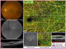
          Optical coherence tomography angiography (OCTA) allows for the evaluation of functional retinal vascular networks without a need for contrast dyes. For sophisticated monitoring and diagnosis of retinal diseases, OCTA capable of providing wide-field and high definition images of retinal vasculature in a single image is desirable. We report OCTA with motion tracking through an auxiliary real-time line scan ophthalmoscope that is clinically feasible to image functional retinal vasculature in patients, with a coverage of more than 60 degrees of retina while still maintaining high definition and resolution. We demonstrate six illustrative cases with unprecedented details of vascular involvement in retinal diseases. In each case, OCTA yields images of the normal and diseased microvasculature at all levels of the retina, with higher resolution than observed with fluorescein angiography. Wide-field OCTA technology will be an important next step in augmenting the utility of OCT technology in clinical practice.

          Related collections

          Most cited references30

          • Record: found
          • Abstract: found
          • Article: not found

          Optical coherence tomography.

          A technique called optical coherence tomography (OCT) has been developed for noninvasive cross-sectional imaging in biological systems. OCT uses low-coherence interferometry to produce a two-dimensional image of optical scattering from internal tissue microstructures in a way that is analogous to ultrasonic pulse-echo imaging. OCT has longitudinal and lateral spatial resolutions of a few micrometers and can detect reflected signals as small as approximately 10(-10) of the incident optical power. Tomographic imaging is demonstrated in vitro in the peripapillary area of the retina and in the coronary artery, two clinically relevant examples that are representative of transparent and turbid media, respectively.
            Bookmark
            • Record: found
            • Abstract: found
            • Article: found
            Is Open Access

            Split-spectrum amplitude-decorrelation angiography with optical coherence tomography

            Amplitude decorrelation measurement is sensitive to transverse flow and immune to phase noise in comparison to Doppler and other phase-based approaches. However, the high axial resolution of OCT makes it very sensitive to the pulsatile bulk motion noise in the axial direction. To overcome this limitation, we developed split-spectrum amplitude-decorrelation angiography (SSADA) to improve the signal-to-noise ratio (SNR) of flow detection. The full OCT spectrum was split into several narrower bands. Inter-B-scan decorrelation was computed using the spectral bands separately and then averaged. The SSADA algorithm was tested on in vivo images of the human macula and optic nerve head. It significantly improved both SNR for flow detection and connectivity of microvascular network when compared to other amplitude-decorrelation algorithms.
              Bookmark
              • Record: found
              • Abstract: found
              • Article: not found

              Idiopathic polypoidal choroidal vasculopathy (IPCV).

              Eleven patients, 40 to 71 years old, had a choroidal vasculopathy that led to hemorrhagic and exudative macular degeneration. The patients had peculiar polypoidal, subretinal, vascular lesions associated with serious and hemorrhagic detachments of the retinal pigment epithelium. This macular disorder, which we have named idiopathic polypoidal choroidal vasculopathy (IPCV), appears to represent a distinct entity that differs clinically and demographically from age-related macular degeneration (AMD) and other macular diseases associated with subretinal neovascularization. Recognition of this condition is important because it may have specific risk factors, natural course, and management considerations that differ from those of age-related macular degeneration.
                Bookmark

                Author and article information

                Journal
                Sci Rep
                Sci Rep
                Scientific Reports
                Nature Publishing Group
                2045-2322
                25 February 2016
                2016
                : 6
                : 22017
                Affiliations
                [1 ]University of Washington, Department of Bioengineering, 3720 15th Ave NE , Seattle, WA 98195, USA
                [2 ]University of Washington, Department of Ophthalmology, 325 Ninth Avenue , Seattle, WA 98104, USA
                [3 ]Carl Zeiss Meditec, Inc. , Dublin, CA 94568, USA
                Author notes
                [*]

                These authors contributed equally to this work.

                Article
                srep22017
                10.1038/srep22017
                4766473
                26912261
                bb60cd75-b912-4abb-9dd2-29f97235f3cd
                Copyright © 2016, Macmillan Publishers Limited

                This work is licensed under a Creative Commons Attribution 4.0 International License. The images or other third party material in this article are included in the article’s Creative Commons license, unless indicated otherwise in the credit line; if the material is not included under the Creative Commons license, users will need to obtain permission from the license holder to reproduce the material. To view a copy of this license, visit http://creativecommons.org/licenses/by/4.0/

                History
                : 13 November 2015
                : 03 February 2016
                Categories
                Article

                Uncategorized
                Uncategorized

                Comments

                Comment on this article