- Record: found
- Abstract: found
- Article: found
A Febrile Infant With Abdominal Erythema and Irritability
research-article

Jaron A. Smith , MD
1 ,
Jennifer Tiller , MD
1 ,
Elizabeth Lagomarsino , MD
2 ,
Joseph Murphy , MD
3
25 March 2023
Read this article at
There is no author summary for this article yet. Authors can add summaries to their articles on ScienceOpen to make them more accessible to a non-specialist audience.
Abstract
Case Report
A 37-day-old male was evaluated in the emergency department for 24 hours of fever
to 102°F, abdominal erythema, and irritability. The patient was also reported to have
10 days of watery, gray stools on an appropriate diet of generic formula and breastmilk.
The patient was the product of a full-term pregnancy and a C-section delivery due
to breech positioning with no perinatal complications.
On initial assessment, the patient was febrile to 38.6°C, tachycardic to 190 bpm,
tachypneic to 70 breaths/minute with normal oxygen saturations and blood pressure.
He was fussy and difficult to console, and he had obvious erythema of the umbilical
and periumbilical region (Figure 1). The abdomen was diffusely tender to palpation
with gross distension. A small umbilical hernia was irreducible with patient straining.
Figure 1.
Physical exam of patient’s abdomen showing periumbilical erythema and mild umbilical
hernia. The patient’s abdomen was also distended and tender to palpation.
Pertinent laboratory findings included a normal complete blood count and complete
metabolic profile, C-reactive protein of 10.7 mg/dL (ref.: 0.0-1.0 mg/dL), and procalcitonin
of 1.47 ng/mL (ref.: <0.05 ng/mL). Urinalysis was unremarkable, urine and blood cultures
were drawn, and an attempted lumbar puncture was unsuccessful.
Plain films demonstrated diffuse gaseous distension of bowel throughout the abdomen
without dilated loops of bowel. Intraluminal gas extended to the rectum. No appreciable
free intraperitoneal air, pneumatosis, or portal venous gas was noted. Abdominal ultrasound
showed hyperemic soft tissue with surrounding inflammatory stranding and a small amount
of fluid with mild hyperemia of the bowel near the site of the herniation suggestive
of an incarcerated umbilical hernia without strangulation. An abdominal computed tomography
(CT) (Figure 2) suggested a central abdominal wall hernia containing loops of small
bowel with associated mild mucosal hyper-enhancement and thickening, potentially concerning
for strangulation/incarceration, but without evidence of bowel obstruction. Alternatively
suggested was a small fluid collection related to an infected umbilicus.
Figure 2.
Axial view of abdominal computed tomography showing hypodense regions in the anterior
abdomen and a small air pocket within those regions.
Piperacillin/tazobactam (80 mg/kg), vancomycin (20 mg/kg), and normal saline (20 mL/kg)
were given in preparation of operative intervention for a presumed incarcerated umbilical
hernia and resulting intra-abdominal infection.
Discussion
Hospital Course
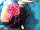
During operative exploration, the periumbilical subcutaneous tissue and surrounding
skin was inflamed and friable, and the umbilical stalk was woody and friable, suggesting
omphalitis. No umbilical hernia with strangulated viscera was identified; however,
an intraperitoneal abscess was found abutting the underside of the anterior abdominal
wall at the base of the umbilical stalk. Extensive debridement of all associated fibrinopurulent
tissues revealed a perforated Meckel’s diverticulum approximately 10 cm proximal to
the ileocecal valve abutting the abscess cavity. A small defect at the base of the
umbilical stalk where the abscess had eroded through the anterior abdominal wall was
repaired. Pathology report would confirm a Meckel’s diverticulum with gastric heterotopia,
transmural inflammation, focal necrosis, and acute serositis.
Postoperative care for sepsis required admission to the pediatric intensive care unit
where the patient was treated with ceftriaxone (50 mg/kg) daily and metronidazole
(10 mg/kg) every 8 hours concurrently for 5 days, then metronidazole alone for an
additional 6 days. Blood and urine cultures had no growth, and abscess cultures were
unable to be obtained. Total parenteral nutrition was required for 7 days, after which
enteral feeds were initiated and well tolerated. The patient was discharged in good
condition. One year from surgery, the patient is growing and developing appropriately.
Discussion of Case and Literature
A ruptured Meckel’s diverticulum in infancy is rare,
1
-4
as is omphalitis occurring outside of the newborn period.
5,6
However, to our knowledge, this is the first reported case of a ruptured Meckel’s
diverticulum leading to omphalitis in infancy outside of the newborn period.
Omphalitis is an infection of the umbilical stump, the umbilicus, and/or the surrounding
tissues, and is distinct from peritonitis, which is infection of the peritoneum, or
abdominal wall.
7
Omphalitis typically presents as a superficial cellulitis. Infection is typically
an external source, associated with cutting of the umbilical cord. Infections are
often polymicrobial, and common pathogens include staphylococcal and streptococcal
species, E. coli, Klebsiella, and anaerobes.
8
These track internally from the severed external umbilical stump, then infect the
nearby soft tissues.
5,7,9
Because there was no guidance provided by culture results in our patient, empiric
therapy was aimed at coverage of these organisms.
Omphalitis can rapidly progress,
5,9
has high morbidity,
5,6,9
and mortality ranges 7% to 15%,
5
thus, early recognition is critical. Because of the cause associated with cutting
and healing of the umbilical cord, it is uncommon for omphalitis to occur outside
the newborn period.
5
-7,10
In these cases, the differential should be broadened, and internal sources that then
infect the umbilical stump and surrounding tissues need to be considered, including
infected remnants of prenatal structures associated with the umbilicus.
10,11
As such, advanced imaging is typically required to identify the source, and surgical
management is often warranted.
5,7
A perforated or ruptured Meckel’s diverticulum is one of these rare causes of nonneonatal
omphalitis. A Meckel’s diverticulum is a true diverticulum that involves all 3 layers
of the bowel wall.
1,2,12
It is a vestigial remnant of the omphalomesenteric duct, also known as the vitelline
duct or yolk stalk.
1,12
Diagnosis of a Meckel’s diverticulum is typically made only when pursued due to clinical
complications, and is challenging since CT and ultrasound may conflate a diverticulum
as normal bowel. A technetium-99m pertechnetate scan is considered the gold standard,
but is not widely available, and is specific only to the Meckel’s diagnosis.
2
Ruptured Meckel’s diverticulum is itself an uncommon complication.
1,4
Although there are case reports and series occurring in the neonatal period
4
and in children older than 12 months,
2,13
the incidence occurring in nonnewborn infancy has only been reported twice since 1975.
1,14
Presenting symptoms are variable, but common complications include bloody stool, bowel
obstruction, sepsis, peritonitis, and much less commonly, as reported in our case,
omphalitis.
1,2,15
Final Diagnosis
Ruptured Meckel’s diverticulum with resulting umbilical stalk abscess and omphalitis.
Conclusion
In summary, omphalitis outside of the neonatal period is rare and requires consideration
of internal causes, including infections and complications of primitive remnants.
Complications of Meckel’s diverticulum are numerous, but less commonly known is perforation,
which can then lead to local and systemic infection, including omphalitis and sepsis.
Surgical consultation was required, and despite advanced imaging, the final diagnosis
was not made until operative management, highlighting the need to involve surgery
early if either of these diagnoses is considered.
Educational Objectives
Omphalitis outside of the neonatal period is a rare entity, and likely to have an
internal cause that must be evaluated with the assistance of imaging and surgical
consultation.
A ruptured Meckel’s diverticulum is a rare complication of a diagnosis that already
requires a high index of suspicion.
Author Contributions
Dr. Smith authored the original draft and all subsequent edits. Drs. Tiller, Lagomarsino,
and Murphy provided mentorship, specialty-specific insight, and reviewed and edited
all drafts.
Related collections
Most cited references15
- Record: found
- Abstract: found
- Article: found
Symptomatic Meckel's Diverticulum in Pediatric Patients—Case Reports and Systematic Review of the Literature
Daniel Keese, Udo Rolle, Stefan Gfroerer … (2019)
- Record: found
- Abstract: found
- Article: not found
Neonatal omphalitis: a review of its serious complications.
Jeremy J. Cusack, Traci Fraser, B Davies (2006)

- Record: found
- Abstract: found
- Article: found
Case report: preoperatively diagnosed perforated Meckel’s diverticulum containing gastric and pancreatic-type mucosa
Georges A. Abizeid, Hager Aref (2017)
Author and article information
Comments
Comment on this article
scite_
0
0
0
0
0
0
0
0
Citing PublicationsSupportingMentioningContrasting
See how this article has been cited at scite.ai
scite shows how a scientific paper has been cited by providing the context of the citation, a classification describing whether it supports, mentions, or contrasts the cited claim, and a label indicating in which section the citation was made.