8
views
0
recommends
+1 Recommend
0 collections
    0
    shares
      • Record: found
      • Abstract: found
      • Article: found
      Is Open Access

      Ki67 index in intrinsic breast cancer subtypes and its association with prognostic parameters

      brief-report

      Read this article at

      Bookmark
          There is no author summary for this article yet. Authors can add summaries to their articles on ScienceOpen to make them more accessible to a non-specialist audience.

          Abstract

          Objectives

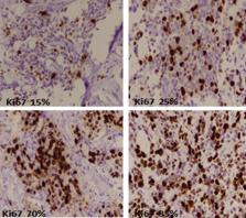
          Ki67 is the most commonly used marker to evaluate proliferative index in breast cancer, however no cutoff values have been clearly defined for high ki67 index. Cancer management should be according to loco-regional profile; therefore, we aimed to determine ki67 index in 1951 cases of intrinsic breast cancer subtypes and its association with other prognostic parameters in our set up.

          Results

          Triple negative breast cancers showed highest ki67 index (mean 50.9 ± 23.7%) followed by Her2neu (mean 42.6 ± 21.6%) and luminal B cancers (mean 34.9 ± 20.05%). Metaplastic and medullary breast cancers significantly showed higher ki67 index as compared to ductal carcinoma, NOS. No significant association of ki67 index was noted with any of the histologic parameters in different subtypes of breast cancer expect for tumor grade. Although, ki67 index is a valuable biomarker in breast cancer, however no independent prognostic significance of ki67 could be established in our study.

          Related collections

          Most cited references16

          • Record: found
          • Abstract: found
          • Article: not found

          Ki-67 as prognostic marker in early breast cancer: a meta-analysis of published studies involving 12 155 patients

          The Ki-67 antigen is used to evaluate the proliferative activity of breast cancer (BC); however, Ki-67's role as a prognostic marker in BC is still undefined. In order to better define the prognostic value of Ki-67/MIB-1, we performed a meta-analysis of studies that evaluated the impact of Ki-67/MIB-1 on disease-free survival (DFS) and/or on overall survival (OS) in early BC. Sixty-eight studies were identified and 46 studies including 12 155 patients were evaluable for our meta-analysis; 38 studies were evaluable for the aggregation of results for DFS, and 35 studies for OS. Patients were considered to present positive tumours for the expression of Ki-67/MIB-1 according to the cut-off points defined by the authors. Ki-67/MIB-1 positivity is associated with higher probability of relapse in all patients (HR=1.93 (95% confidence interval (CI): 1.74–2.14); P<0.001), in node-negative patients (HR=2.31 (95% CI: 1.83–2.92); P<0.001) and in node-positive patients (HR=1.59 (95% CI: 1.35–1.87); P<0.001). Furthermore, Ki-67/MIB-1 positivity is associated with worse survival in all patients (HR=1.95 (95% CI: 1.70–2.24; P<0.001)), node-negative patients (HR=2.54 (95% CI: 1.65–3.91); P<0.001) and node-positive patients (HR=2.33 (95% CI: 1.83–2.95); P<0.001). Our meta-analysis suggests that Ki-67/MIB-1 positivity confers a higher risk of relapse and a worse survival in patients with early BC.
            Bookmark
            • Record: found
            • Abstract: found
            • Article: not found

            Proliferation marker Ki-67 in early breast cancer.

            Molecular markers have been extensively investigated with a view to providing early and accurate information on long-term outcome and prediction of response to treatment of early breast cancer. Proliferation is a key feature of the progression of tumors and is now widely estimated by the immunohistochemical assessment of the nuclear antigen Ki-67. The expression of Ki-67 correlates with other measurements of proliferation, including S-phase and bromodeoxyuridine uptake. High Ki-67 is a sign of poor prognosis associated with a good chance of clinical response to chemotherapy, but its independent significance is modest and does not merit measurements in most routine clinical scenarios. However, its application as a pharmacodynamic intermediate marker of the effectiveness of medical therapy holds great promise for rapid evaluation of new drugs.
              Bookmark
              • Record: found
              • Abstract: found
              • Article: not found

              Estrogen receptor analyses. Correlation of biochemical and immunohistochemical methods using monoclonal antireceptor antibodies.

              Attempts at histochemical localization of estrogen receptor with anti-steroid antibody or some fluoresceinated estrogens have given unacceptable sensitivities and specificities when compared with biochemical methods or clinical response. In the present study a monoclonal antibody against estrogen receptor (H222 Sp gamma) was used on cryostat sections of freshly frozen breast tumors with a peroxidase-antiperoxidase immunoperoxidase technique. Biochemical receptor analyses were by dextran-coated charcoal analyses. Tumors from three separate cohorts of patients were studied as follows: population A, 62 primary breast cancers from 1983; population B, 72 primary lesions stored from 1976 to 1983; and population C, 23 patients with metastases, treated with hormonal therapy. Distinct staining was seen in the cell nucleus. A semiquantitative relationship was seen between histochemical score assessment of staining and biochemical assay in each cohort. The sensitivity and specificity using a threshold of 75 for the histochemical score and more than 20 femtomoles/mg of protein for dextran-coated charcoal analyses were as follows: population A, specificity, 89%, and sensitivity, 95%; population B, specificity, 94%, and sensitivity 88%; and for population C, the comparison was with objective clinical response yielding specificity, 89%, and sensitivity, 93%.
                Bookmark

                Author and article information

                Contributors
                doc_atif2005@yahoo.com
                drkhashmi72@gmail.com
                irfanzafar892@gmail.com
                smk.state.alchemist@gmail.com
                Muhammad_edhi@brown.edu
                javaria_29@hotmail.com
                huzaifaurwah@gmail.com
                huda.ha16@gmail.com
                Naveen.faridi@gmail.com
                dramirkhan04@gmail.com
                Journal
                BMC Res Notes
                BMC Res Notes
                BMC Research Notes
                BioMed Central (London )
                1756-0500
                23 September 2019
                23 September 2019
                2019
                : 12
                : 605
                Affiliations
                [1 ]ISNI 0000 0004 0637 9066, GRID grid.415915.d, Liaquat National Hospital and Medical College, ; Karachi, Pakistan
                [2 ]ISNI 0000 0004 0609 1628, GRID grid.416335.6, Nishtar Medical College Hospital, ; Multan, Pakistan
                [3 ]ISNI 0000 0004 1936 9094, GRID grid.40263.33, Brown University, ; Providence, RI USA
                [4 ]CMH Institute of Medical Sciences, Multan, Pakistan
                [5 ]GRID grid.440459.8, Kandahar University, ; Kandahar, 3802 Afghanistan
                Author information
                http://orcid.org/0000-0002-8763-7690
                Article
                4653
                10.1186/s13104-019-4653-x
                6755684
                31547858
                bafc7263-5351-4627-a5e4-61ab15d93d82
                © The Author(s) 2019

                Open AccessThis article is distributed under the terms of the Creative Commons Attribution 4.0 International License ( http://creativecommons.org/licenses/by/4.0/), which permits unrestricted use, distribution, and reproduction in any medium, provided you give appropriate credit to the original author(s) and the source, provide a link to the Creative Commons license, and indicate if changes were made. The Creative Commons Public Domain Dedication waiver ( http://creativecommons.org/publicdomain/zero/1.0/) applies to the data made available in this article, unless otherwise stated.

                History
                : 5 February 2019
                : 17 September 2019
                Categories
                Research Note
                Custom metadata
                © The Author(s) 2019

                Medicine
                breast cancer,intrinsic subtypes of breast cancer,ki67 index,er,pr,her2neu
                Medicine
                breast cancer, intrinsic subtypes of breast cancer, ki67 index, er, pr, her2neu

                Comments

                Comment on this article