7
views
0
recommends
+1 Recommend
0 collections
    0
    shares
      • Record: found
      • Abstract: found
      • Article: found
      Is Open Access

      Long-term results of MyoRing treatment of keratoconus Translated title: Resultados a largo plazo del tratamiento del queratocono con MyoRing

      research-article
      a , b , * , c , d
      Journal of Optometry
      Elsevier
      MyoRing, Keratoconus, ICRS, CISIS, MyoRing, Queratocono, ICRS, CISIS

      Read this article at

      Bookmark
          There is no author summary for this article yet. Authors can add summaries to their articles on ScienceOpen to make them more accessible to a non-specialist audience.

          Abstract

          Purpose

          To study long-term results of MyoRing treatment of keratoconus.

          Methods

          Retrospective study of MyoRing implantation into a corneal pocket for keratoconus.

          Results

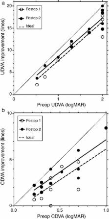
          Corneal thickness at the thinnest point remained unchanged, SIM K's, manifest sphere and cylinder were significantly improved at the first follow-up 9 months postoperatively and remained stable until the last follow-up about 5 years after surgery. Uncorrected and corrected distance visual acuity (UDVA, CDVA) were significantly improved at the first follow-up 9 months postoperatively and were further ameliorated until the last follow-up about 5 years after surgery.

          Conclusion

          The treatment was safe and effective with continuing improvement of visual acuity during the 5 years after surgery.

          Resumen

          Objetivo

          Estudiar los resultados a largo plazo del tratamiento del queratocono con MyoRing.

          Método

          Estudio retrospectivo de la implantación del MyoRing en un bolsillo corneal en los casos de queratocono.

          Resultados

          El espesor de la córnea en el punto más fino no reflejó cambios, mejorando los valores de SIM K, esfera y cilindro manifiesto durante el seguimiento a los 9 meses de la intervención y permaneciendo entonces estables hasta el último seguimiento a los 5 años de la misma. La agudeza visual no corregida y corregida (UDVA, CDVA) mejoró considerablemente a los 9 meses de la intervención, y continuaron mejorando significativamente hasta el último seguimiento a los 5 años de la misma.

          Conclusión

          El tratamiento resultó seguro y efectivo, con mejora continua de la agudeza visual durante los 5 años posteriores a la cirugía.

          Related collections

          Most cited references19

          • Record: found
          • Abstract: found
          • Article: not found

          Intracorneal rings for keratoconus and keratectasia.

          Intrastromal corneal ring segments were designed to achieve refractive adjustment by flattening the cornea. Recently, they have been used to reshape keratoconic corneas to improve uncorrected visual acuity, best corrected visual acuity, and contact lens tolerance and to delay or prevent the need for keratoplasty. Intracorneal ring segments have several distinct and important advantages. New thicknesses and different ring sizes and the use of femtosecond lasers to dissect channels inside the cornea will likely improve the surgical outcomes. This article reviews the latest data published or presented at meetings on the correction of keratoconus and keratectasia by intracorneal ring segments.
            Bookmark
            • Record: found
            • Abstract: found
            • Article: not found

            Progression of keratoconus by longitudinal assessment with corneal topography.

            To investigate the longitudinal changes in corneal topographic indices over time in patients with mild keratoconus (KC) and to determine predictive factors for the increase in corneal curvature.
              Bookmark
              • Record: found
              • Abstract: found
              • Article: not found

              Outcome analysis of intracorneal ring segments for the treatment of keratoconus based on visual, refractive, and aberrometric impairment.

              To analyze the outcomes of intracorneal ring segment (ICRS) implantation for the treatment of keratoconus based on preoperative visual impairment.
                Bookmark

                Author and article information

                Contributors
                Journal
                J Optom
                J Optom
                Journal of Optometry
                Elsevier
                1888-4296
                1989-1342
                02 March 2016
                Apr-Jun 2017
                02 March 2016
                : 10
                : 2
                : 123-129
                Affiliations
                [a ]Gutsehen Eye Center, Ybbs/Vienna, Austria
                [b ]Department of Ophthalmology, Medical University, Innsbruck, Austria
                [c ]Department of Ophthalmology, Medical University, St. Pölten, Austria
                [d ]Department of Ophthalmology, Hospital Barmherzige Schwestern, Ried, Austria
                Author notes
                [* ]Corresponding author at: Stauwerkstrasse 1, A-3370 Ybbs, Austria. daxer@ 123456gutsehen.at
                Article
                S1888-4296(16)00004-2
                10.1016/j.optom.2016.01.002
                5383460
                26922840
                b9e5d203-91ec-46d3-af51-6677e87c756a
                © 2016 Spanish General Council of Optometry. Published by Elsevier Espa˜na, S.L.U.

                This is an open access article under the CC BY-NC-ND license (http://creativecommons.org/licenses/by-nc-nd/4.0/).

                History
                : 29 November 2015
                : 11 January 2016
                Categories
                Original Article

                myoring,keratoconus,icrs,cisis,queratocono
                myoring, keratoconus, icrs, cisis, queratocono

                Comments

                Comment on this article