32
views
0
recommends
+1 Recommend
1 collections
    1
    shares
      • Record: found
      • Abstract: found
      • Article: found
      Is Open Access

      Investigating Klebsiella pneumoniae biofilm preservation for scanning electron microscopy

      methods-article

      Read this article at

      ScienceOpenPublisherPMC
      Bookmark
          There is no author summary for this article yet. Authors can add summaries to their articles on ScienceOpen to make them more accessible to a non-specialist audience.

          Abstract

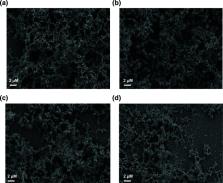
          Klebsiella pneumoniae biofilm formation is associated with chronic and relapsing infections. Scanning electron microscopy (SEM) is a powerful tool for characterizing biofilm structure and studying their formation. Reliable visualization of biofilm structure requires careful sample preservation, otherwise there may be loss of non-covalent interactions that are susceptible to damage during the dehydration and washing preparation steps. However, no standard procedure has been adopted in the literature to fix K. pneumoniae biofilm for scanning electron microscopy studies. This lack of standardization makes it challenging to compare results between studies and determine the degree to which native structures have been preserved. To advance this critical area of study, we investigated different scanning electron microscopy fixation methods for K. pneumoniae biofilm preservation. Our study reveals the impact preparation steps can have on retaining in biofilm architecture observed using scanning electron microscopy. Using fixation methods developed through our studies, we show that although species that overproduce capsular extracellular polysaccharides produced more robust biofilms, K. pneumoniae can form a developed biofilm in the absence of capsular polysaccharides.

          Related collections

          Most cited references38

          • Record: found
          • Abstract: found
          • Article: not found

          The biofilm matrix.

          The microorganisms in biofilms live in a self-produced matrix of hydrated extracellular polymeric substances (EPS) that form their immediate environment. EPS are mainly polysaccharides, proteins, nucleic acids and lipids; they provide the mechanical stability of biofilms, mediate their adhesion to surfaces and form a cohesive, three-dimensional polymer network that interconnects and transiently immobilizes biofilm cells. In addition, the biofilm matrix acts as an external digestive system by keeping extracellular enzymes close to the cells, enabling them to metabolize dissolved, colloidal and solid biopolymers. Here we describe the functions, properties and constituents of the EPS matrix that make biofilms the most successful forms of life on earth.
            Bookmark
            • Record: found
            • Abstract: found
            • Article: not found

            Physiological heterogeneity in biofilms.

            Biofilms contain bacterial cells that are in a wide range of physiological states. Within a biofilm population, cells with diverse genotypes and phenotypes that express distinct metabolic pathways, stress responses and other specific biological activities are juxtaposed. The mechanisms that contribute to this genetic and physiological heterogeneity include microscale chemical gradients, adaptation to local environmental conditions, stochastic gene expression and the genotypic variation that occurs through mutation and selection. Here, we discuss the processes that generate chemical gradients in biofilms, the genetic and physiological responses of the bacteria as they adapt to these gradients and the techniques that can be used to visualize and measure the microscale physiological heterogeneities of bacteria in biofilms.
              Bookmark
              • Record: found
              • Abstract: found
              • Article: found
              Is Open Access

              Hypervirulent (hypermucoviscous) Klebsiella pneumoniae

              A new hypervirulent (hypermucoviscous) variant of Klebsiella pneumoniae has emerged. First described in the Asian Pacific Rim, it now increasingly recognized in Western countries. Defining clinical features are the ability to cause serious, life-threatening community-acquired infection in younger healthy hosts, including liver abscess, pneumonia, meningitis and endophthalmitis and the ability to metastatically spread, an unusual feature for enteric Gram-negative bacilli in the non-immunocompromised. Despite infecting a healthier population, significant morbidity and mortality occurs. Although epidemiologic features are still being defined, colonization, particularly intestinal colonization, appears to be a critical step leading to infection. However the route of entry remains unclear. The majority of cases described to date are in Asians, raising the issue of a genetic predisposition vs. geospecific strain acquisition. The traits that enhance its virulence when compared with “classical” K. pneumoniae are the ability to more efficiently acquire iron and perhaps an increase in capsule production, which confers the hypermucoviscous phenotype. An objective diagnostic test suitable for routine use in the clinical microbiology laboratory is needed. If/when these strains become increasingly resistant to antimicrobials, we will be faced with a frightening clinical scenario.
                Bookmark

                Author and article information

                Journal
                Access Microbiol
                Access Microbiol
                acmi
                acmi
                Access Microbiology
                Microbiology Society
                2516-8290
                2023
                3 February 2023
                3 February 2023
                : 5
                : 2
                : 000470.v3
                Affiliations
                [ 1] Department of Molecular Biosciences, The University of Texas at Austin , Austin, TX 78712, USA
                [ 2] Center for Biomedical Research Support, The University of Texas at Austin , Austin, TX 78712, USA
                [ 3] Center for Systems and Synthetic Biology, The University of Texas at Austin , Austin, TX 78712, USA
                [ 4] John Ring LaMontagne Center for Infectious Diseases, The University of Texas at Austin , Austin, TX 78712, USA
                [ ]Present address: Division of Immunity and Pathogenesis, Burnett School of Biomedical Sciences, College of Medicine, University of Central Florida, Orlando, FL 32837, USA
                Author notes
                *Correspondence: Bryan W. Davies, bwdavies@ 123456utexas.edu
                *Correspondence: Renee M. Fleeman, renee.fleeman@ 123456ucf.edu
                Author information
                https://orcid.org/0000-0001-7103-461X
                https://orcid.org/0000-0002-2046-0341
                Article
                000470.v3
                10.1099/acmi.0.000470.v3
                9996182
                36910511
                b942d602-0b2e-4f2a-9a66-9277dc350014
                © 2023 The Authors

                This is an open-access article distributed under the terms of the Creative Commons Attribution License.

                History
                : 13 July 2022
                : 02 January 2023
                Funding
                Funded by: National Institute of Allergy and Infectious Diseases
                Award ID: K99AI163295
                Award Recipient : ReneeM Fleeman
                Funded by: National Institute of Allergy and Infectious Diseases
                Award ID: AI159203
                Award Recipient : BryanW Davies
                Funded by: National Institute of Allergy and Infectious Diseases
                Award ID: AI148419
                Award Recipient : BryanW Davies
                Funded by: National Institute of Allergy and Infectious Diseases
                Award ID: AI125337
                Award Recipient : BryanW Davies
                Categories
                Methods
                Custom metadata
                0
                0

                biofilm,klebsiella pneumoniae,scanning electron microscopy

                Comments

                Comment on this article