83
views
0
recommends
+1 Recommend
0 collections
    0
    shares
      • Record: found
      • Abstract: found
      • Article: found
      Is Open Access

      Non-small cell lung cancer cells survived ionizing radiation treatment display cancer stem cell and epithelial-mesenchymal transition phenotypes

      research-article

      Read this article at

      Bookmark
          There is no author summary for this article yet. Authors can add summaries to their articles on ScienceOpen to make them more accessible to a non-specialist audience.

          Abstract

          Ionizing radiation (IR) is used for patients diagnosed with unresectable non small cell lung cancer (NSCLC), however radiotherapy remains largely palliative due to radioresistance. Cancer stem cells (CSCs), as well as epithelial-mesenchymal transition (EMT), may contribute to drug and radiation resistance mechanisms in solid tumors. Here we investigated the molecular phenotype of A549 and H460 NSCLC cells that survived treatment with IR (5Gy) and are growing as floating tumor spheres and cells that are maintained in a monolayer after irradiation.

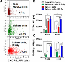
          Non-irradiated and irradiated cells were collected after one week, seeded onto ultra low attachment plates and propagated as tumor spheres. Bulk NSCLC cells which survived radiation and grew in spheres express cancer stem cell surface and embryonic stem cell markers and are able to self-renew, and generate differentiated progeny. These cells also have a mesenchymal phenotype. Particularly, the radiation survived sphere cells express significantly higher levels of CSC markers (CD24 and CD44), nuclear β-catenin and EMT markers (Snail1, Vimentin, and N-cadherin) than non-irradiated lung tumor sphere cells. Upregulated levels of Oct-4, Sox2 and beta-catenin were detected in H460 cells maintained in a monolayer after irradiation, but not in radiation survived adherent A459 cells.

          PDGFR-beta was upregulated in radiation survived sphere cells and in radiation survived adherent cells in both A549 and H460 cell lines. Combining IR treatment with axitinib or dasatinib, inhibitors with anti-PDFGR activity, potentiates the efficacy of NSCLC radiotherapy in vitro.

          Our findings suggest that radiation survived cells have a complex phenotype combining the properties of CSCs and EMT. CD44, SNAIL and PDGFR-beta are dramatically upregulated in radiation survived cells and might be considered as markers of radiotherapy response in NSCLC.

          Related collections

          Most cited references47

          • Record: found
          • Abstract: found
          • Article: not found

          Highly tumorigenic lung cancer CD133+ cells display stem-like features and are spared by cisplatin treatment.

          The identification of lung tumor-initiating cells and associated markers may be useful for optimization of therapeutic approaches and for predictive and prognostic information in lung cancer patients. CD133, a surface glycoprotein linked to organ-specific stem cells, was described as a marker of cancer-initiating cells in different tumor types. Here, we report that a CD133+, epithelial-specific antigen-positive (CD133+ESA+) population is increased in primary nonsmall cell lung cancer (NSCLC) compared with normal lung tissue and has higher tumorigenic potential in SCID mice and expression of genes involved in stemness, adhesion, motility, and drug efflux than the CD133(-) counterpart. Cisplatin treatment of lung cancer cells in vitro resulted in enrichment of CD133+ fraction both after acute cytotoxic exposure and in cells with stable cisplatin-resistant phenotype. Subpopulations of CD133+ABCG2+ and CD133+CXCR4+ cells were spared by in vivo cisplatin treatment of lung tumor xenografts established from primary tumors. A tendency toward shorter progression-free survival was observed in CD133+ NSCLC patients treated with platinum-containing regimens. Our results indicate that chemoresistant populations with highly tumorigenic and stem-like features are present in lung tumors. The molecular features of these cells may provide the rationale for more specific therapeutic targeting and the definition of predictive factors in clinical management of this lethal disease.
            Bookmark
            • Record: found
            • Abstract: found
            • Article: not found

            Aldehyde dehydrogenase 1 is a tumor stem cell-associated marker in lung cancer.

            Tumor contains small population of cancer stem cells (CSC) that are responsible for its maintenance and relapse. Analysis of these CSCs may lead to effective prognostic and therapeutic strategies for the treatment of cancer patients. We report here the identification of CSCs from human lung cancer cells using Aldefluor assay followed by fluorescence-activated cell sorting analysis. Isolated cancer cells with relatively high aldehyde dehydrogenase 1 (ALDH1) activity display in vitro features of CSCs, including capacities for proliferation, self-renewal, and differentiation, resistance to chemotherapy, and expressing CSC surface marker CD133. In vivo experiments show that the ALDH1-positive cells could generate tumors that recapitulate the heterogeneity of the parental cancer cells. Immunohistochemical analysis of 303 clinical specimens from three independent cohorts of lung cancer patients and controls show that expression of ALDH1 is positively correlated with the stage and grade of lung tumors and related to a poor prognosis for the patients with early-stage lung cancer. ALDH1 is therefore a lung tumor stem cell-associated marker. These findings offer an important new tool for the study of lung CSCs and provide a potential prognostic factor and therapeutic target for treatment of the patients with lung cancer.
              Bookmark
              • Record: found
              • Abstract: found
              • Article: not found

              The hypoxic microenvironment maintains glioblastoma stem cells and promotes reprogramming towards a cancer stem cell phenotype.

              Glioblastomas are highly lethal cancers that contain cellular hierarchies with self-renewing cancer stem cells that can propagate tumors in secondary transplant assays. The potential significance of cancer stem cells in cancer biology has been demonstrated by studies showing contributions to therapeutic resistance, angiogenesis and tumor dispersal. We recently reported that physiologic oxygen levels differentially induce hypoxia inducible factor-2alpha (HIF2alpha) levels in cancer stem cells. HIF1alpha functioned in proliferation and survival of all cancer cells but also was activated in normal neural progenitors suggesting a potentially restricted therapeutic index while HIF2alpha was essential in only in cancer stem cells and was not expressed by normal neural progenitors demonstrating HIF2alpha is a cancer stem cell specific target. We now extend these studies to examine the role of hypoxia in regulating tumor cell plasticity. We find that hypoxia promotes the self-renewal capability of the stem and non-stem population as well as promoting a more stem-like phenotype in the non-stem population with increased neurosphere formation as well as upregulation of important stem cell factors, such as OCT4, NANOG and c-MYC. The importance of HIF2alpha was further supported as forced expression of non-degradable HIF2alpha induced a cancer stem cell marker and augmented the tumorigenic potential of the non-stem population. This novel finding may indicate a specific role of HIF2alpha in promoting glioma tumorigenesis. The unexpected plasticity of the non-stem glioma population and the stem-like phenotype emphasizes the importance of developing therapeutic strategies targeting the microenvironmental influence on the tumor in addition to cancer stem cells.
                Bookmark

                Author and article information

                Journal
                Mol Cancer
                Mol. Cancer
                Molecular Cancer
                BioMed Central
                1476-4598
                2013
                16 August 2013
                : 12
                : 94
                Affiliations
                [1 ]University of Pittsburgh Cancer Institute, Pittsburgh, PA 15213, USA
                [2 ]Department of Medicine, Pittsburg, PA 15213, USA
                [3 ]Departments of Pathology, University of Pittsburgh, Pittsburgh, PA 15213, USA
                [4 ]University of Pittsburgh Cancer Institute, Hillman Cancer Center, Rm. 1.19c, 5117 Center Ave, Pittsburgh, PA 15213, USA
                Article
                1476-4598-12-94
                10.1186/1476-4598-12-94
                3751356
                23947765
                b8d12837-796f-4078-8fd6-889c34bfb2e2
                Copyright ©2013 Gomez-casal et al.; licensee BioMed Central Ltd.

                This is an Open Access article distributed under the terms of the Creative Commons Attribution License ( http://creativecommons.org/licenses/by/2.0), which permits unrestricted use, distribution, and reproduction in any medium, provided the original work is properly cited.

                History
                : 3 April 2013
                : 14 August 2013
                Categories
                Research

                Oncology & Radiotherapy
                non-small cell lung cancer,ionizing radiation,cancer stem cell,epithelial-mesenchymal transition

                Comments

                Comment on this article