0
views
0
recommends
+1 Recommend
0 collections
    0
    shares
      • Record: found
      • Abstract: found
      • Article: found
      Is Open Access

      Longitudinal MRI in the context of in utero surgery for open spina bifida: A descriptive study

      research-article

      Read this article at

      Bookmark
          There is no author summary for this article yet. Authors can add summaries to their articles on ScienceOpen to make them more accessible to a non-specialist audience.

          Abstract

          Introduction

          Fetal surgery for open spina bifida (OSB) requires comprehensive preoperative assessment using imaging for appropriate patient selection and to evaluate postoperative efficacy and complications. We explored patient access and conduct of fetal magnetic resonance imaging (MRI) for prenatal assessment of OSB patients eligible for fetal surgery. We compared imaging acquisition and reporting to the International Society of Ultrasound in Obstetrics and Gynecology MRI performance guidelines.

          Material and methods

          We surveyed access to fetal MRI for OSB in referring fetal medicine units (FMUs) in the UK and Ireland, and two NHS England specialist commissioned fetal surgery centers (FSCs) at University College London Hospital, and University Hospitals KU Leuven Belgium. To study MRI acquisition protocols, we retrospectively analyzed fetal MRI images before and after fetal surgery for OSB.

          Results

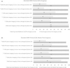
          MRI for fetal OSB was accessible with appropriate specialists available to supervise, perform, and report scans. The average time to arrange a fetal MRI appointment from request was 4 ± 3 days (range, 0–10), the average scan time available was 37 ± 16 min (range, 20–80 min), with 15 ± 11 min (range, 0–30 min) extra time to repeat sequences as required. Specific MRI acquisition protocols, and MRI reporting templates were available in only 32% and 18% of units, respectively. Satisfactory T2‐weighted (T2W) brain imaging acquired in three orthogonal planes was achieved preoperatively in all centers, and 6 weeks postoperatively in 96% of FSCs and 78% of referring FMUs. However, for T2W spine image acquisition referring FMUs were less able to provide three orthogonal planes presurgery (98% FSC vs. 50% FMU, p < 0.001), and 6 weeks post‐surgery (100% FSC vs. 48% FMU, p < 0.001). Other standard imaging recommendations such as T1‐weighted (T1W), gradient echo (GE) or echoplanar fetal brain and spine imaging in one or two orthogonal planes were more likely available in FSCs compared to FMUs pre‐ and post‐surgery ( p < 0.001).

          Conclusions

          There was timely access to supervised MRI for OSB fetal surgery assessment. However, the provision of images of the fetal brain and spine in sufficient orthogonal planes, which are required for determining eligibility and to determine the reversal of hindbrain herniation after fetal surgery, were less frequently acquired. Our evidence suggests the need for specific guidance in relation to fetal MRI for OSB. We propose an example guidance for MRI acquisition and reporting.

          Abstract

          Fetal surgery for open spina bifida (OSB) is performed in selected patients. We explored ease of access, acquisition and reporting of fetal magnetic resonance imaging (MRI) in patients undergoing fetal surgery for OSB. Agreed criteria for fetal MRI in cases of OSB is a priority to ensure correct patient selection as well as to critically evaluate the efficacy of fetal surgery.

          Related collections

          Most cited references45

          • Record: found
          • Abstract: found
          • Article: not found

          A randomized trial of prenatal versus postnatal repair of myelomeningocele.

          Prenatal repair of myelomeningocele, the most common form of spina bifida, may result in better neurologic function than repair deferred until after delivery. We compared outcomes of in utero repair with standard postnatal repair. We randomly assigned eligible women to undergo either prenatal surgery before 26 weeks of gestation or standard postnatal repair. One primary outcome was a composite of fetal or neonatal death or the need for placement of a cerebrospinal fluid shunt by the age of 12 months. Another primary outcome at 30 months was a composite of mental development and motor function. The trial was stopped for efficacy of prenatal surgery after the recruitment of 183 of a planned 200 patients. This report is based on results in 158 patients whose children were evaluated at 12 months. The first primary outcome occurred in 68% of the infants in the prenatal-surgery group and in 98% of those in the postnatal-surgery group (relative risk, 0.70; 97.7% confidence interval [CI], 0.58 to 0.84; P<0.001). Actual rates of shunt placement were 40% in the prenatal-surgery group and 82% in the postnatal-surgery group (relative risk, 0.48; 97.7% CI, 0.36 to 0.64; P<0.001). Prenatal surgery also resulted in improvement in the composite score for mental development and motor function at 30 months (P=0.007) and in improvement in several secondary outcomes, including hindbrain herniation by 12 months and ambulation by 30 months. However, prenatal surgery was associated with an increased risk of preterm delivery and uterine dehiscence at delivery. Prenatal surgery for myelomeningocele reduced the need for shunting and improved motor outcomes at 30 months but was associated with maternal and fetal risks. (Funded by the National Institutes of Health; ClinicalTrials.gov number, NCT00060606.).
            Bookmark
            • Record: found
            • Abstract: found
            • Article: not found

            Basic principles of diffusion-weighted imaging.

            In diffusion-weighted MRI (DWI), image contrast is determined by the random microscopic motion of water protons. During the last years, DWI has become an important modality in the diagnostic work-up of acute ischemia in the CNS. There are also a few promising reports about the application of DWI to other regions in the human body, such as the vertebral column or the abdomen. This manuscript provides an introduction into the basics of DWI and Diffusion Tensor imaging. The potential of various MR sequences in concert with diffusion preparation are discussed with respect to acquisition speed, spatial resolution, and sensitivity to bulk physiologic motion. More advanced diffusion measurement techniques, such as high angular resolution diffusion imaging, are also addressed. Copyright 2002 Elsevier Science Ireland, Ltd.
              Bookmark
              • Record: found
              • Abstract: found
              • Article: not found

              Laminar organization of the human fetal cerebrum revealed by histochemical markers and magnetic resonance imaging.

              The developing human cerebrum displays age-specific changes in its patterns of lamination. Among these, the subplate zone is the most prominent transient compartment because growing major afferent systems temporarily reside in this zone, establish synapses and take part in cellular interactions that are crucial for subsequent cortical development. We explored the potential of magnetic resonance imaging (MRI) for tracing the developmental history of the most prominent cortical layer (the subplate zone) and other laminar compartments of the fetal cerebral wall between 15 and 36 weeks post-ovulation. We found that changes in the MRI lamination pattern of the human fetal cerebral wall are predominantly caused by changes in the subplate zone. Histochemical staining of the extracellular matrix (ECM) enables selective visualization of the subplate zone and correlation with an increase in MRI signal intensity in the subplate zone and ingrowth and accumulation of thalamocortical and corticocortical afferents and their subsequent relocation to the cortical plate. Thus, dynamic changes in the MRI appearance of the subplate zone and histochemical staining of its ECM can be used as indirect parameters for an assessment of normal versus disturbed unfolding of crucial histogenetic events that are involved in prenatal shaping of the human cerebral cortex.
                Bookmark

                Author and article information

                Contributors
                n.mufti@ucl.ac.uk
                Journal
                Acta Obstet Gynecol Scand
                Acta Obstet Gynecol Scand
                10.1111/(ISSN)1600-0412
                AOGS
                Acta Obstetricia et Gynecologica Scandinavica
                John Wiley and Sons Inc. (Hoboken )
                0001-6349
                1600-0412
                20 November 2023
                February 2024
                : 103
                : 2 ( doiID: 10.1111/aogs.v103.2 )
                : 322-333
                Affiliations
                [ 1 ] Elizabeth Garrett Anderson Institute for Women's Health University College London London UK
                [ 2 ] School of Biomedical Engineering and Imaging Sciences (BMEIS) King's College London London UK
                [ 3 ] Department of Radiology University Hospitals Katholieke Universiteit (KU) Leuven Belgium
                [ 4 ] Pediatric Neurosurgery Department Great Ormond Street Hospital for Children London UK
                [ 5 ] Department of Neurosurgery University Hospitals Katholieke Universiteit (KU) Leuven Belgium
                [ 6 ] Department of Obstetrics and Gynecology University Hospitals Katholieke Universiteit (KU) Leuven Belgium
                [ 7 ] Medical Physics and Biomedical Engineering University College London London UK
                Author notes
                [*] [* ] Correspondence

                Nada Mufti, Elizabeth Garrett Anderson Institute for Women's Health, University College London, 1st Floor Charles Bell House, 43‐45 Foley Street, London W1W 7TS, UK.

                Email: n.mufti@ 123456ucl.ac.uk

                Author information
                https://orcid.org/0000-0001-9839-8085
                https://orcid.org/0000-0002-4920-945X
                Article
                AOGS14711 AOGS-23-0477.R1
                10.1111/aogs.14711
                10823411
                37984808
                b8952d33-b0d2-44be-845f-1e24dfd7fdd3
                © 2023 The Authors. Acta Obstetricia et Gynecologica Scandinavica published by John Wiley & Sons Ltd on behalf of Nordic Federation of Societies of Obstetrics and Gynecology (NFOG).

                This is an open access article under the terms of the http://creativecommons.org/licenses/by-nc-nd/4.0/ License, which permits use and distribution in any medium, provided the original work is properly cited, the use is non‐commercial and no modifications or adaptations are made.

                History
                : 17 September 2023
                : 16 June 2023
                : 17 October 2023
                Page count
                Figures: 6, Tables: 3, Pages: 12, Words: 6566
                Funding
                Funded by: Engineering and Physical Sciences Research Council , doi 10.13039/501100000266;
                Award ID: NS/A000049/1
                Award ID: NS/A000050/1
                Award ID: NS/A000027/1
                Award ID: EP/L016
                Funded by: Wellcome Trust , doi 10.13039/100010269;
                Award ID: 203148/Z/16/Z
                Award ID: 203145Z/16/Z
                Award ID: WT101957
                Categories
                Original Research Article
                Fetal Medicine
                Custom metadata
                2.0
                February 2024
                Converter:WILEY_ML3GV2_TO_JATSPMC version:6.3.6 mode:remove_FC converted:29.01.2024

                Obstetrics & Gynecology
                accessibility,fetal surgery,magnetic resonance imaging,open spina bifida,protocols,sequences

                Comments

                Comment on this article