8
views
0
recommends
+1 Recommend
0 collections
    0
    shares
      • Record: found
      • Abstract: found
      • Article: found
      Is Open Access

      Astrocyte calcium dysfunction causes early network hyperactivity in Alzheimer’s disease

      research-article

      Read this article at

      Bookmark
          There is no author summary for this article yet. Authors can add summaries to their articles on ScienceOpen to make them more accessible to a non-specialist audience.

          Summary

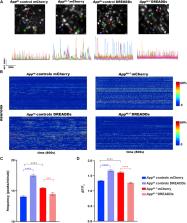
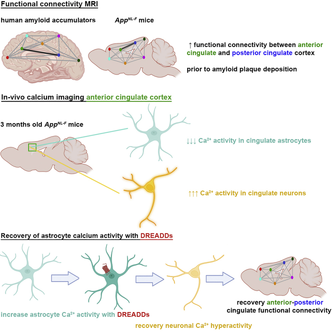
          Dysfunctions of network activity and functional connectivity (FC) represent early events in Alzheimer’s disease (AD), but the underlying mechanisms remain unclear. Astrocytes regulate local neuronal activity in the healthy brain, but their involvement in early network hyperactivity in AD is unknown. We show increased FC in the human cingulate cortex several years before amyloid deposition. We find the same early cingulate FC disruption and neuronal hyperactivity in App NL-F mice. Crucially, these network disruptions are accompanied by decreased astrocyte calcium signaling. Recovery of astrocytic calcium activity normalizes neuronal hyperactivity and FC, as well as seizure susceptibility and day/night behavioral disruptions. In conclusion, we show that astrocytes mediate initial features of AD and drive clinically relevant phenotypes.

          Graphical abstract

          Highlights

          • The cingulate cortex of humans and mice shows early functional deficits in AD

          • Astrocyte calcium signaling is decreased before the presence of amyloid plaques

          • Recovery of astrocyte calcium signals mitigates neuronal hyperactivity

          • Recovery of astrocytes normalizes cingulate connectivity and behavior disruptions

          Abstract

          Shah et al. show disrupted cingulate functional connectivity in mice and humans before amyloid deposition. Cingulate astrocytes demonstrate decreased calcium signaling, while neurons are hyperactive. Restoring the regulatory activity of astrocytes recovers neuronal hyperactivity, cingulate connectivity, and behavior deficits, suggesting that astrocytes represent a major player in early Alzheimer’s disease.

          Related collections

          Most cited references90

          • Record: found
          • Abstract: found
          • Article: not found

          Automated anatomical labeling of activations in SPM using a macroscopic anatomical parcellation of the MNI MRI single-subject brain.

          An anatomical parcellation of the spatially normalized single-subject high-resolution T1 volume provided by the Montreal Neurological Institute (MNI) (D. L. Collins et al., 1998, Trans. Med. Imag. 17, 463-468) was performed. The MNI single-subject main sulci were first delineated and further used as landmarks for the 3D definition of 45 anatomical volumes of interest (AVOI) in each hemisphere. This procedure was performed using a dedicated software which allowed a 3D following of the sulci course on the edited brain. Regions of interest were then drawn manually with the same software every 2 mm on the axial slices of the high-resolution MNI single subject. The 90 AVOI were reconstructed and assigned a label. Using this parcellation method, three procedures to perform the automated anatomical labeling of functional studies are proposed: (1) labeling of an extremum defined by a set of coordinates, (2) percentage of voxels belonging to each of the AVOI intersected by a sphere centered by a set of coordinates, and (3) percentage of voxels belonging to each of the AVOI intersected by an activated cluster. An interface with the Statistical Parametric Mapping package (SPM, J. Ashburner and K. J. Friston, 1999, Hum. Brain Mapp. 7, 254-266) is provided as a freeware to researchers of the neuroimaging community. We believe that this tool is an improvement for the macroscopical labeling of activated area compared to labeling assessed using the Talairach atlas brain in which deformations are well known. However, this tool does not alleviate the need for more sophisticated labeling strategies based on anatomical or cytoarchitectonic probabilistic maps.
            Bookmark
            • Record: found
            • Abstract: found
            • Article: found
            Is Open Access

            The neuropathological diagnosis of Alzheimer’s disease

            Alzheimer’s disease is a progressive neurodegenerative disease most often associated with memory deficits and cognitive decline, although less common clinical presentations are increasingly recognized. The cardinal pathological features of the disease have been known for more than one hundred years, and today the presence of these amyloid plaques and neurofibrillary tangles are still required for a pathological diagnosis. Alzheimer’s disease is the most common cause of dementia globally. There remain no effective treatment options for the great majority of patients, and the primary causes of the disease are unknown except in a small number of familial cases driven by genetic mutations. Confounding efforts to develop effective diagnostic tools and disease-modifying therapies is the realization that Alzheimer’s disease is a mixed proteinopathy (amyloid and tau) frequently associated with other age-related processes such as cerebrovascular disease and Lewy body disease. Defining the relationships between and interdependence of various co-pathologies remains an active area of investigation. This review outlines etiologically-linked pathologic features of Alzheimer’s disease, as well as those that are inevitable findings of uncertain significance, such as granulovacuolar degeneration and Hirano bodies. Other disease processes that are frequent, but not inevitable, are also discussed, including pathologic processes that can clinically mimic Alzheimer’s disease. These include cerebrovascular disease, Lewy body disease, TDP-43 proteinopathies and argyrophilic grain disease. The purpose of this review is to provide an overview of Alzheimer’s disease pathology, its defining pathologic substrates and the related pathologies that can affect diagnosis and treatment. Electronic supplementary material The online version of this article (10.1186/s13024-019-0333-5) contains supplementary material, which is available to authorized users.
              Bookmark
              • Record: found
              • Abstract: found
              • Article: not found

              DREADDs for Neuroscientists.

              Bryan Roth (2016)
              To understand brain function, it is essential that we discover how cellular signaling specifies normal and pathological brain function. In this regard, chemogenetic technologies represent valuable platforms for manipulating neuronal and non-neuronal signal transduction in a cell-type-specific fashion in freely moving animals. Designer Receptors Exclusively Activated by Designer Drugs (DREADD)-based chemogenetic tools are now commonly used by neuroscientists to identify the circuitry and cellular signals that specify behavior, perceptions, emotions, innate drives, and motor functions in species ranging from flies to nonhuman primates. Here I provide a primer on DREADDs highlighting key technical and conceptual considerations and identify challenges for chemogenetics going forward.
                Bookmark

                Author and article information

                Contributors
                Journal
                Cell Rep
                Cell Rep
                Cell Reports
                Cell Press
                2211-1247
                23 August 2022
                23 August 2022
                23 August 2022
                : 40
                : 8
                : 111280
                Affiliations
                [1 ]Laboratory for the Research of Neurodegenerative Diseases, VIB Center for Brain and Disease Research, KU Leuven, 3000 Leuven, Belgium
                [2 ]Biomedical MRI, Department of Imaging and Pathology, KU Leuven, 3000 Leuven, Belgium
                [3 ]Laboratory of Glia Biology, VIB Center for Brain and Disease Research, KU Leuven, 3000 Leuven, Belgium
                [4 ]Laboratory for Cognitive Neurology, Department of Neurosciences, Leuven Brain Institute (LBI), KU Leuven, 3000 Leuven, Belgium
                [5 ]Neuro-electronics Research Flanders, 3000 Leuven, Belgium
                [6 ]Laboratory for Neuropathology, Department of Imaging and Pathology, LBI, KU Leuven, 3000 Leuven, Belgium
                [7 ]Laboratory of Biological Psychology, KU-Leuven, 3000 Leuven, Belgium
                [8 ]UK Dementia Research Institute at University College London, WC1E 6BT London, UK
                Author notes
                []Corresponding author disha.shah@ 123456kuleuven.be
                [∗∗ ]Corresponding author b.destrooper@ 123456ucl.ac.uk
                [9]

                Lead contact

                Article
                S2211-1247(22)01100-7 111280
                10.1016/j.celrep.2022.111280
                9433881
                36001964
                b83be1bc-5e8a-4492-aa30-7979503a1ad6
                © 2022 The Authors.

                This is an open access article under the CC BY-NC-ND license (http://creativecommons.org/licenses/by-nc-nd/4.0/).

                History
                : 17 May 2022
                : 30 June 2022
                : 5 August 2022
                Categories
                Article

                Cell biology
                astrocytes,amyloid pathology,bold rsfmri,calcium signaling,neuronal hyperactivity,app mice

                Comments

                Comment on this article