73
views
0
recommends
+1 Recommend
0 collections
    0
    shares
      • Record: found
      • Abstract: found
      • Article: found
      Is Open Access

      Susceptibility-Weighted Imaging of the Anatomic Variation of Thalamostriate Vein and Its Tributaries

      research-article

      Read this article at

      Bookmark
          There is no author summary for this article yet. Authors can add summaries to their articles on ScienceOpen to make them more accessible to a non-specialist audience.

          Abstract

          Background and Purpose

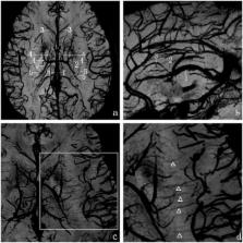
          Thalamostriate vein (TSV) is an important tributary of the internal cerebral vein, which mainly drains the basal ganglia and deep medulla. The purpose of this study was to explore the anatomic variation and quality of TSV and its smaller tributaries using susceptibility-weighted imaging (SWI).

          Methods

          We acquired SWI images in 40 volunteers on a 3.0T MR system using an 8-channel high-resolution phased array coil. The frequencies of the TSV and its tributaries were evaluated. We classified TSV into types I (forming a venous angle) and II (forming a false venous angle). We classified anterior caudate vein (ACV)into types 1 (1 trunk) and 2 (2 trunks) as well as into types A (joiningTSV), B (joining anterior septal vein), and C (joining the angle of both veins).

          Results

          The TSV drains the areas of caudate nucleus, internal capsule,lentiform nucleus, external capsule, claustrum, extreme capsule and the white matter of the frontoparietal lobes,except thalamus. The frequencies of the TSV, ACV and transverse caudate vein (ACV) were 92.5%, 87.5% and 63.8%, respectively. We found TSV types I and II in 79.7%, and 20.3% with significantly different constitution ratios ( P< 0.05). The most common types of ACV were type 1 (90.0%) and type A (64.3%).

          Conclusion

          The complex three-dimensional (3D) venous architecture of TSV and its small tributaries manifests great variation, with significant and practical implications for neurosurgery.

          Related collections

          Most cited references26

          • Record: found
          • Abstract: found
          • Article: not found

          Small vessels in the human brain: MR venography with deoxyhemoglobin as an intrinsic contrast agent.

          To assess a magnetic resonance (MR) imaging method for depicting small veins in the brain, a three-dimensional, long echo time, gradient-echo sequence that depended on the paramagnetic property of deoxyhemoglobin was used. Veins with diameters smaller than a pixel were depicted. This MR imaging method is easy to implement and may prove helpful in the evaluation of venous diseases.
            Bookmark
            • Record: found
            • Abstract: found
            • Article: not found

            Susceptibility weighted imaging at ultra high magnetic field strengths: theoretical considerations and experimental results.

            We present numerical simulations and experimental results for susceptibility weighted imaging (SWI) at 7 T. Magnitude, phase, and SWI contrast were simulated for different voxel geometries and imaging parameters, resulting in an echo time of 14 msec for optimum contrast between veins and surrounding tissue. Slice thickness of twice the in-plane voxel size or more resulted in optimum vessel visibility. Phantom and in vivo data are in very good agreement with the simulations and the delineation of vessels at 7 T was superior compared to lower field strengths. The phase of the complex data reveals anatomical details that are complementary to the corresponding magnitude images. Susceptibility weighted imaging at very high field strengths is a promising technique because of its high sensitivity to tissue susceptibility, its low specific absorption rate, and the phase's negligible sensitivity to B(1) inhomogeneities.
              Bookmark
              • Record: found
              • Abstract: found
              • Article: not found

              Nonnvasive assessment of vascular architecture and function during modulated blood oxygenation using susceptibility weighted magnetic resonance imaging.

              Susceptibility weighted imaging (SWI) is a BOLD-sensitive method for visualizing anatomical features such as small cerebral veins in high detail. The purpose of this study was to evaluate high-resolution SWI in combination with a modulation of blood oxygenation by breathing of air, carbogen, and oxygen and to directly visualize the effects of changing blood oxygenation on the magnetic field inside and around venous blood vessels. Signal changes associated with the response to carbogen and oxygen breathing were evaluated in different anatomic regions in healthy volunteers and in two patients with brain tumors. In the magnitude images inhalation of carbogen led to significant signal intensity changes ranging from +4.4 +/- 1.9% to +9.5 +/- 1.4% in gray matter and no significant changes in thalamus, putamen, and white matter. During oxygen breathing mean signal changes were smaller than during carbogen breathing. The method is capable of producing high-resolution functional maps of BOLD response to carbogen and oxygen breathing as well as high-resolution images of venous vasculature. Its sensitivity to changes in blood oxygenation was demonstrated by in vivo visualization of the BOLD effect via phase imaging.
                Bookmark

                Author and article information

                Contributors
                Role: Editor
                Journal
                PLoS One
                PLoS ONE
                plos
                plosone
                PLoS ONE
                Public Library of Science (San Francisco, CA USA )
                1932-6203
                27 October 2015
                2015
                : 10
                : 10
                : e0141513
                Affiliations
                [1 ]Department of Human Anatomy, Wenzhou Medical University, Wenzhou, Zhejiang, China
                [2 ]Department of Radiology, the 1 st Affiliated Hospital of Wenzhou Medical University, Wenzhou, Zhejiang, China
                [3 ]Department of Neurosurgery, the 2 nd Affiliated Hospital of Wenzhou Medical University, Wenzhou, Zhejiang, China
                Shenzhen institutes of advanced technology, CHINA
                Author notes

                Competing Interests: The authors have declared that no competing interests exist.

                Conceived and designed the experiments: X-FZ J-CL C-CC. Performed the experiments: X-FZ J-CL X-DW. Analyzed the data: X-FZ C-GR MC. Contributed reagents/materials/analysis tools: X-FZ J-CL C-GR. Wrote the paper: X-FZ J-CL X-DW C-CC.

                Article
                PONE-D-15-27668
                10.1371/journal.pone.0141513
                4624633
                26506095
                b81180aa-082d-45a4-b13d-13537ecda112
                Copyright @ 2015

                This is an open access article distributed under the terms of the Creative Commons Attribution License, which permits unrestricted use, distribution, and reproduction in any medium, provided the original author and source are credited

                History
                : 30 June 2015
                : 7 October 2015
                Page count
                Figures: 7, Tables: 3, Pages: 11
                Funding
                This work was supported by the Natural Science Foundation of Zhejiang Province, China (NO. LY15C110001).
                Categories
                Research Article
                Custom metadata
                All relevant data are within the paper.

                Uncategorized
                Uncategorized

                Comments

                Comment on this article