0
views
0
recommends
+1 Recommend
0 collections
    0
    shares
      • Record: found
      • Abstract: found
      • Article: found
      Is Open Access

      Tumoral periprostatic adipose tissue exovesicles-derived miR-20a-5p regulates prostate cancer cell proliferation and inflammation through the RORA gene

      research-article

      Read this article at

      Bookmark
          There is no author summary for this article yet. Authors can add summaries to their articles on ScienceOpen to make them more accessible to a non-specialist audience.

          Abstract

          Background

          From the first steps of prostate cancer (PCa) initiation, tumours are in contact with the most-proximal adipose tissue called periprostatic adipose tissue (PPAT). Extracellular vesicles are important carriers of non-coding RNA such as miRNAs that are crucial for cellular communication. The secretion of extracellular vesicles by PPAT may play a key role in the interactions between adipocytes and tumour. Analysing the PPAT exovesicles (EVs) derived-miRNA content can be of great relevance for understanding tumour progression and aggressiveness.

          Methods

          A total of 24 samples of human PPAT and 17 samples of perivesical adipose tissue (PVAT) were used. EVs were characterized by western blot and transmission electron microscopy (TEM), and uptake by PCa cells was verified by confocal microscopy. PPAT and PVAT explants were cultured overnight, EVs were isolated, and miRNA content expression profile was analysed. Pathway and functional enrichment analyses were performed seeking potential miRNA targets. In vitro functional studies were evaluated using PCa cells lines, miRNA inhibitors and target gene silencers.

          Results

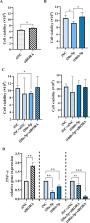
          Western blot and TEM revealed the characteristics of EVs derived from PPAT (PPAT-EVs) samples. The EVs were up taken and found in the cytoplasm of PCa cells. Nine miRNAs were differentially expressed between PPAT and PVAT samples. The RORA gene (RAR Related Orphan Receptor A) was identified as a common target of 9 miRNA-regulated pathways. In vitro functional analysis revealed that the RORA gene was regulated by PPAT-EVs-derived miRNAs and was found to be implicated in cell proliferation and inflammation.

          Conclusion

          Tumour periprostatic adipose tissue is linked to PCa tumour aggressiveness and could be envisaged for new therapeutic strategies.

          Supplementary Information

          The online version contains supplementary material available at 10.1186/s12967-024-05458-3.

          Related collections

          Most cited references19

          • Record: found
          • Abstract: found
          • Article: found
          Is Open Access

          Biological properties of extracellular vesicles and their physiological functions

          In the past decade, extracellular vesicles (EVs) have been recognized as potent vehicles of intercellular communication, both in prokaryotes and eukaryotes. This is due to their capacity to transfer proteins, lipids and nucleic acids, thereby influencing various physiological and pathological functions of both recipient and parent cells. While intensive investigation has targeted the role of EVs in different pathological processes, for example, in cancer and autoimmune diseases, the EV-mediated maintenance of homeostasis and the regulation of physiological functions have remained less explored. Here, we provide a comprehensive overview of the current understanding of the physiological roles of EVs, which has been written by crowd-sourcing, drawing on the unique EV expertise of academia-based scientists, clinicians and industry based in 27 European countries, the United States and Australia. This review is intended to be of relevance to both researchers already working on EV biology and to newcomers who will encounter this universal cell biological system. Therefore, here we address the molecular contents and functions of EVs in various tissues and body fluids from cell systems to organs. We also review the physiological mechanisms of EVs in bacteria, lower eukaryotes and plants to highlight the functional uniformity of this emerging communication system.
            Bookmark
            • Record: found
            • Abstract: found
            • Article: not found

            The 2014 International Society of Urological Pathology (ISUP) Consensus Conference on Gleason Grading of Prostatic Carcinoma: Definition of Grading Patterns and Proposal for a New Grading System.

            In November, 2014, 65 prostate cancer pathology experts, along with 17 clinicians including urologists, radiation oncologists, and medical oncologists from 19 different countries gathered in a consensus conference to update the grading of prostate cancer, last revised in 2005. The major conclusions were: (1) Cribriform glands should be assigned a Gleason pattern 4, regardless of morphology; (2) Glomeruloid glands should be assigned a Gleason pattern 4, regardless of morphology; (3) Grading of mucinous carcinoma of the prostate should be based on its underlying growth pattern rather than grading them all as pattern 4; and (4) Intraductal carcinoma of the prostate without invasive carcinoma should not be assigned a Gleason grade and a comment as to its invariable association with aggressive prostate cancer should be made. Regarding morphologies of Gleason patterns, there was clear consensus on: (1) Gleason pattern 4 includes cribriform, fused, and poorly formed glands; (2) The term hypernephromatoid cancer should not be used; (3) For a diagnosis of Gleason pattern 4, it needs to be seen at 10x lens magnification; (4) Occasional/seemingly poorly formed or fused glands between well-formed glands is insufficient for a diagnosis of pattern 4; (5) In cases with borderline morphology between Gleason pattern 3 and pattern 4 and crush artifacts, the lower grade should be favored; (6) Branched glands are allowed in Gleason pattern 3; (7) Small solid cylinders represent Gleason pattern 5; (8) Solid medium to large nests with rosette-like spaces should be considered to represent Gleason pattern 5; and (9) Presence of unequivocal comedonecrosis, even if focal is indicative of Gleason pattern 5. It was recognized by both pathologists and clinicians that despite the above changes, there were deficiencies with the Gleason system. The Gleason grading system ranges from 2 to 10, yet 6 is the lowest score currently assigned. When patients are told that they have a Gleason score 6 out of 10, it implies that their prognosis is intermediate and contributes to their fear of having a more aggressive cancer. Also, in the literature and for therapeutic purposes, various scores have been incorrectly grouped together with the assumption that they have a similar prognosis. For example, many classification systems consider Gleason score 7 as a single score without distinguishing 3+4 versus 4+3, despite studies showing significantly worse prognosis for the latter. The basis for a new grading system was proposed in 2013 by one of the authors (J.I.E.) based on data from Johns Hopkins Hospital resulting in 5 prognostically distinct Grade Groups. This new system was validated in a multi-institutional study of over 20,000 radical prostatectomy specimens, over 16,000 needle biopsy specimens, and over 5,000 biopsies followed by radiation therapy. There was broad (90%) consensus for the adoption of this new prostate cancer Grading system in the 2014 consensus conference based on: (1) the new classification provided more accurate stratification of tumors than the current system; (2) the classification simplified the number of grading categories from Gleason scores 2 to 10, with even more permutations based on different pattern combinations, to Grade Groups 1 to 5; (3) the lowest grade is 1 not 6 as in Gleason, with the potential to reduce overtreatment of indolent cancer; and (4) the current modified Gleason grading, which forms the basis for the new grade groups, bears little resemblance to the original Gleason system. The new grades would, for the foreseeable future, be used in conjunction with the Gleason system [ie. Gleason score 3+3=6 (Grade Group 1)]. The new grading system and the terminology Grade Groups 1-5 have also been accepted by the World Health Organization for the 2016 edition of Pathology and Genetics: Tumours of the Urinary System and Male Genital Organs.
              Bookmark
              • Record: found
              • Abstract: found
              • Article: not found

              Adipocyte Exosomes Promote Melanoma Aggressiveness through Fatty Acid Oxidation: A Novel Mechanism Linking Obesity and Cancer.

              Malignant progression results from a dynamic cross-talk between stromal and cancer cells. Recent evidence suggests that this cross-talk is mediated to a significant extent by exosomes, nanovesicles secreted by most cell types and which allow the transfer of proteins, lipids, and nucleic acids between cells. Adipocytes are a major component of several tumor microenvironments, including that of invasive melanoma, where cells have migrated to the adipocyte-rich hypodermic layer of the skin. We show that adipocytes secrete exosomes in abundance, which are then taken up by tumor cells, leading to increased migration and invasion. Using mass spectrometry, we analyzed the proteome of adipocyte exosomes. Interestingly, these vesicles carry proteins implicated in fatty acid oxidation (FAO), a feature highly specific to adipocyte exosomes. We further show that, in the presence of adipocyte exosomes, FAO is increased in melanoma cells. Inhibition of this metabolic pathway completely abrogates the exosome-mediated increase in migration. Moreover, in obese mice and humans, both the number of exosomes secreted by adipocytes as well as their effect on FAO-dependent cell migration are amplified. These observations might in part explain why obese melanoma patients have a poorer prognosis than their nonobese counterparts. Cancer Res; 76(14); 4051-7. ©2016 AACR.
                Bookmark

                Author and article information

                Contributors
                mrch2424@gmail.com , matilde.rodriguez@iispv.cat
                Journal
                J Transl Med
                J Transl Med
                Journal of Translational Medicine
                BioMed Central (London )
                1479-5876
                15 July 2024
                15 July 2024
                2024
                : 22
                : 661
                Affiliations
                [1 ]Disease Biomarkers and Molecular Mechanisms Group. IISPV. Joan, XXIII University Hospital, Universitat Rovira i Virgili, ( https://ror.org/00g5sqv46) Tarragona, Spain
                [2 ]GRID grid.411435.6, ISNI 0000 0004 1767 4677, Urology Unit, , Joan XXIII University Hospital, ; Tarragona, Spain
                [3 ]GRID grid.411435.6, ISNI 0000 0004 1767 4677, Pathology Unit, , Joan XXIII University Hospital, ; Tarragona, Spain
                [4 ]GRID grid.411435.6, ISNI 0000 0004 1767 4677, Institut d’Investigació Sanitària Pere Virgili. Hospital Universitari de Tarragona Joan XXIII, ; C/ Dr. Mallafré Guasch, 4, Tarragona, 43007 Spain
                Author information
                http://orcid.org/0000-0003-1531-2165
                Article
                5458
                10.1186/s12967-024-05458-3
                11251289
                39010137
                b77ed071-c5dc-41ba-aca3-4a0394fb4a79
                © The Author(s) 2024

                Open Access This article is licensed under a Creative Commons Attribution 4.0 International License, which permits use, sharing, adaptation, distribution and reproduction in any medium or format, as long as you give appropriate credit to the original author(s) and the source, provide a link to the Creative Commons licence, and indicate if changes were made. The images or other third party material in this article are included in the article’s Creative Commons licence, unless indicated otherwise in a credit line to the material. If material is not included in the article’s Creative Commons licence and your intended use is not permitted by statutory regulation or exceeds the permitted use, you will need to obtain permission directly from the copyright holder. To view a copy of this licence, visit http://creativecommons.org/licenses/by/4.0/. The Creative Commons Public Domain Dedication waiver ( http://creativecommons.org/publicdomain/zero/1.0/) applies to the data made available in this article, unless otherwise stated in a credit line to the data.

                History
                : 2 May 2024
                : 29 June 2024
                Funding
                Funded by: FundRef http://dx.doi.org/10.13039/501100004587, Instituto de Salud Carlos III;
                Award ID: PI17/00877
                Award ID: PI20/00418
                Award ID: PI24/00015
                Funded by: FundRef http://dx.doi.org/10.13039/501100004837, Ministerio de Ciencia e Innovación;
                Award ID: PID2023-146128OB-I00
                Funded by: FundRef http://dx.doi.org/10.13039/501100002704, Fundación Científica Asociación Española Contra el Cáncer;
                Award ID: PRYES246650RODR
                Categories
                Research
                Custom metadata
                © BioMed Central Ltd., part of Springer Nature 2024

                Medicine
                periprostatic adipose tissue,exovesicles,mirnas,prostate cancer,rora gene
                Medicine
                periprostatic adipose tissue, exovesicles, mirnas, prostate cancer, rora gene

                Comments

                Comment on this article