17
views
0
recommends
+1 Recommend
0 collections
    0
    shares
      • Record: found
      • Abstract: found
      • Article: found
      Is Open Access

      Identification of a novel cathelicidin antimicrobial peptide from ducks and determination of its functional activity and antibacterial mechanism

      research-article

      Read this article at

      Bookmark
          There is no author summary for this article yet. Authors can add summaries to their articles on ScienceOpen to make them more accessible to a non-specialist audience.

          Abstract

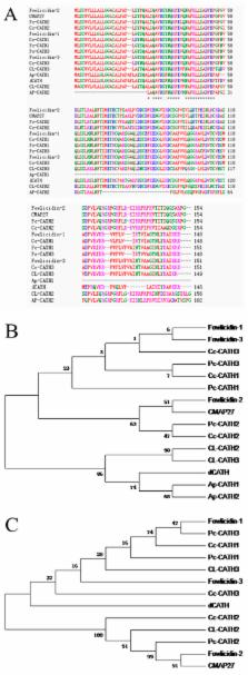
          The family of antimicrobial peptide, cathelicidins, which plays important roles against infections in animals, has been identified from many species. Here, we identified a novel avian cathelicidin ortholog from ducks and named dCATH. The cDNA sequence of dCATH encodes a predicted 146-amino-acid polypeptide composed of a 17-residue signal peptide, a 109-residue conserved cathelin domain and a 20-residue mature peptide. Phylogenetic analysis demonstrated that dCATH is highly divergent from other avian peptides. The α-helical structure of the peptide exerted strong antimicrobial activity against a broad range of bacteria in vitro, with most minimum inhibitory concentrations in the range of 2 to 4 μM. Moreover, dCATH also showed cytotoxicity, lysing 50% of mammalian erythrocytes in the presence or absence of 10% fetal calf serum at concentrations of 32 μM or 20 μM and killing 50% HaCaT cells at a concentration of 10 μM. The effects on bacterial outer and inner membranes, as examined by scanning electron microscope and transmission electron microscopy, indicate that dCATH kills microbial cells by increasing permeability, causing a loss of membrane integrity.

          Related collections

          Most cited references33

          • Record: found
          • Abstract: found
          • Article: not found

          Human cathelicidin, hCAP-18, is processed to the antimicrobial peptide LL-37 by extracellular cleavage with proteinase 3.

          Cathelicidins are a family of antimicrobial proteins found in the peroxidase-negative granules of neutrophils. The known biologic functions reside in the C-terminus, which must be cleaved from the holoprotein to become active. Bovine and porcine cathelicidins are cleaved by elastase from the azurophil granules to yield the active antimicrobial peptides. The aim of this study was to identify the physiological setting for cleavage of the only human cathelicidin, hCAP-18, to liberate the antibacterial and cytotoxic peptide LL-37 and to identify the protease responsible for this cleavage. Immunoelectron microscopy demonstrated that both hCAP-18 and azurophil granule proteins were present in the phagolysosome. Immunoblotting revealed no detectable cleavage of hCAP-18 in cells after phagocytosis. In contrast, hCAP-18 was cleaved to generate LL-37 in exocytosed material. Of the 3 known serine proteases from azurophil granules, proteinase 3 was solely responsible for cleavage of hCAP-18 after exocytosis. This is the first detailed study describing the generation of a human antimicrobial peptide from a promicrobicidal protein, and it demonstrates that the generation of active antimicrobial peptides from common proproteins occurs differently in related species. (Blood. 2001;97:3951-3959)
            Bookmark
            • Record: found
            • Abstract: found
            • Article: not found

            Mechanism of action of the antimicrobial peptide buforin II: buforin II kills microorganisms by penetrating the cell membrane and inhibiting cellular functions.

            The mechanism of action of buforin II, which is a 21-amino acid peptide with a potent antimicrobial activity against a broad range of microorganisms, was studied using fluorescein isothiocyanate (FITC)-labeled buforin II and a gel-retardation experiment. Its mechanism of action was compared with that of the well-characterized magainin 2, which has a pore-forming activity on the cell membrane. Buforin II killed Esche-richia coli without lysing the cell membrane even at 5 times minimal inhibitory concentration (MIC) at which buforin II reduced the viable cell numbers by 6 orders of magnitude. However, magainin 2 lysed the cell to death under the same condition. FITC-labeled buforin II was found to penetrate the cell membrane and accumulate inside E. coli even below its MIC, whereas FITC-labeled magainin 2 remained outside or on the cell wall even at its MIC. The gel-retardation experiment showed that buforin II bound to DNA and RNA of the cells over 20 times strongly than magainin 2. All these results indicate that buforin II inhibits the cellular functions by binding to DNA and RNA of cells after penetrating the cell membranes, resulting in the rapid cell death, which is quite different from that of magainin 2 even though they are structurally similar: a linear amphipathic alpha-helical peptide.
              Bookmark
              • Record: found
              • Abstract: found
              • Article: not found

              The duck genome and transcriptome provide insight into an avian influenza virus reservoir species

              The duck (Anas platyrhynchos) is one of the principal natural hosts of influenza A viruses. We present the duck genome sequence and perform deep transcriptome analyses to investigate immune-related genes. Our data indicate that the duck possesses a contractive immune gene repertoire, as in chicken and zebra finch, and this repertoire has been shaped through lineage-specific duplications. We identify genes that are responsive to influenza A viruses using the lung transcriptomes of control ducks and ones that were infected with either a highly pathogenic (A/duck/Hubei/49/05) or a weakly pathogenic (A/goose/Hubei/65/05) H5N1 virus. Further, we show how the duck’s defense mechanisms against influenza infection have been optimized through the diversification of its β-defensin and butyrophilin-like repertoires. These analyses, in combination with the genomic and transcriptomic data, provide a resource for characterizing the interaction between host and influenza viruses.
                Bookmark

                Author and article information

                Journal
                Sci Rep
                Sci Rep
                Scientific Reports
                Nature Publishing Group
                2045-2322
                26 November 2015
                2015
                : 5
                : 17260
                Affiliations
                [1 ]College of Animal Science and Technology, Northeast Agricultural University , Harbin 150030, P.R. China
                Author notes
                Article
                srep17260
                10.1038/srep17260
                4660463
                26608073
                b736a8f2-a717-4485-ba40-7bc6635fa021
                Copyright © 2015, Macmillan Publishers Limited

                This work is licensed under a Creative Commons Attribution 4.0 International License. The images or other third party material in this article are included in the article’s Creative Commons license, unless indicated otherwise in the credit line; if the material is not included under the Creative Commons license, users will need to obtain permission from the license holder to reproduce the material. To view a copy of this license, visit http://creativecommons.org/licenses/by/4.0/

                History
                : 27 July 2015
                : 27 October 2015
                Categories
                Article

                Uncategorized
                Uncategorized

                Comments

                Comment on this article