1
views
0
recommends
+1 Recommend
0 collections
    0
    shares
      • Record: found
      • Abstract: found
      • Article: found
      Is Open Access

      Automated analysis of computerized morphological features of cell clusters associated with malignancy on bile duct brushing whole slide images

      research-article

      Read this article at

      Bookmark
          There is no author summary for this article yet. Authors can add summaries to their articles on ScienceOpen to make them more accessible to a non-specialist audience.

          Abstract

          Background

          Bile duct brush specimens are difficult to interpret as they often present inflammatory and reactive backgrounds due to the local effects of stricture, atypical reactive changes, or previously installed stents, and often have low to intermediate cellularity. As a result, diagnosis of biliary adenocarcinomas is challenging and often results in large interobserver variability and low sensitivity

          Objective

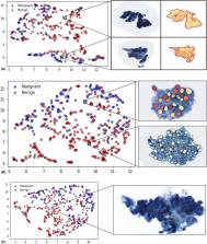
          In this work, we used computational image analysis to evaluate the role of nuclear morphological and texture features of epithelial cell clusters to predict the presence of pancreatic and biliary tract adenocarcinoma on digitized brush cytology specimens.

          Methods

          Whole slide images from 124 patients, either diagnosed as benign or malignant based on clinicopathological correlation, were collected and randomly split into training ( S T, N = 58) and testing ( S v , N = 66) sets, with the exception of cases diagnosed as atypical on cytology were included in S v . Nuclear boundaries on cell clusters extracted from each image were segmented via a watershed algorithm. A total of 536 quantitative morphometric features pertaining to nuclear shape, size, and aggregate cluster texture were extracted from within the cell clusters. The most predictive features from patients in S T were selected via rank‐sum, t‐test, and minimum redundancy maximum relevance (mRMR) schemes. The selected features were then used to train three machine‐learning classifiers.

          Results

          Malignant clusters tended to exhibit lower textural homogeneity within the nucleus, greater textural entropy around the nuclear membrane, and longer minor axis lengths. The sensitivity of cytology alone was 74% (without atypicals) and 46% (with atypicals). With machine diagnosis, the sensitivity improved to 68% from 46% when atypicals were included and treated as nonmalignant false negatives. The specificity of our model was 100% within the atypical category.

          Conclusion

          We achieved an area under the receiver operating characteristic curve (AUC) of 0.79 on S v , which included atypical cytological diagnosis.

          Abstract

          Pancreatobiliary adenocarcinomas can be diagnosed with a computerized approach with sensitivity of 63%. Atypical cases can be diagnosed with 100% specificity.

          Related collections

          Most cited references30

          • Record: found
          • Abstract: found
          • Article: found
          Is Open Access

          UMAP: Uniform Manifold Approximation and Projection

            Bookmark
            • Record: found
            • Abstract: not found
            • Article: not found

            On a Test of Whether one of Two Random Variables is Stochastically Larger than the Other

              Bookmark
              • Record: found
              • Abstract: found
              • Article: not found

              Feature selection based on mutual information: criteria of max-dependency, max-relevance, and min-redundancy.

              Feature selection is an important problem for pattern classification systems. We study how to select good features according to the maximal statistical dependency criterion based on mutual information. Because of the difficulty in directly implementing the maximal dependency condition, we first derive an equivalent form, called minimal-redundancy-maximal-relevance criterion (mRMR), for first-order incremental feature selection. Then, we present a two-stage feature selection algorithm by combining mRMR and other more sophisticated feature selectors (e.g., wrappers). This allows us to select a compact set of superior features at very low cost. We perform extensive experimental comparison of our algorithm and other methods using three different classifiers (naive Bayes, support vector machine, and linear discriminate analysis) and four different data sets (handwritten digits, arrhythmia, NCI cancer cell lines, and lymphoma tissues). The results confirm that mRMR leads to promising improvement on feature selection and classification accuracy.
                Bookmark

                Author and article information

                Contributors
                axm788@case.edu
                Journal
                Cancer Med
                Cancer Med
                10.1002/(ISSN)2045-7634
                CAM4
                Cancer Medicine
                John Wiley and Sons Inc. (Hoboken )
                2045-7634
                24 October 2022
                March 2023
                : 12
                : 5 ( doiID: 10.1002/cam4.v12.5 )
                : 6365-6378
                Affiliations
                [ 1 ] Department of Biomedical Engineering Case Western Reserve University Cleveland Ohio USA
                [ 2 ] Department of Pathology Case Western Reserve University School of Medicine, University Hospitals Cleveland Medical Center Cleveland Ohio USA
                [ 3 ] Louis Stokes Cleveland Veterans Administration Medical Center Cleveland Ohio USA
                Author notes
                [*] [* ] Correspondence

                Anant Madabhushi, Department of Biomedical Engineering, Case Western Reserve University, Cleveland, OH, USA.

                Email: axm788@ 123456case.edu

                Author information
                https://orcid.org/0000-0003-0719-4007
                Article
                CAM45365 CAM4-2022-01-0467.R1
                10.1002/cam4.5365
                10028025
                36281473
                b703716c-58e9-42cf-98c0-a0bad95e7117
                © 2022 The Authors. Cancer Medicine published by John Wiley & Sons Ltd.

                This is an open access article under the terms of the http://creativecommons.org/licenses/by/4.0/ License, which permits use, distribution and reproduction in any medium, provided the original work is properly cited.

                History
                : 01 June 2022
                : 30 March 2022
                : 07 August 2022
                Page count
                Figures: 7, Tables: 2, Pages: 14, Words: 7561
                Funding
                Funded by: National Cancer Institute , doi 10.13039/100000054;
                Award ID: 1U54CA254566‐01
                Award ID: 1U01CA248226‐01
                Award ID: 1U01CA239055‐01
                Award ID: R01CA257612‐01A1
                Award ID: R01CA220581‐01A1
                Award ID: R01CA216579‐01A1
                Award ID: R01CA208236‐01A1
                Award ID: R01CA202752‐01A1
                Award ID: R01CA249992‐01A1
                Funded by: National Heart, Lung and Blood Institute
                Award ID: R01HL15807101A1
                Award ID: 1R01HL15127701A1
                Funded by: National Institute of Biomedical Imaging and Bioengineering , doi 10.13039/100000070;
                Award ID: 1R43EB028736‐01
                Funded by: National Center for Research Resources , doi 10.13039/100000097;
                Award ID: 1C06 RR12463‐01
                Funded by: United States Department of Veterans Affairs Biomedical Laboratory Research and Development Service
                Award ID: IBX004121A
                Funded by: Breast Cancer Research Program
                Award ID: W81XWH‐19‐1‐0668
                Funded by: Prostate Cancer Research Program , doi 10.13039/100014039;
                Award ID: W81XWH‐20‐1‐0851
                Award ID: W81XWH‐15‐1‐0558
                Funded by: Lung Cancer Research Program
                Award ID: W81XWH‐20‐1‐0595
                Award ID: W81XWH‐18‐1‐0440
                Funded by: Peer Reviewed Cancer Research Program , doi 10.13039/100014042;
                Award ID: W81XWH‐21‐1‐0345
                Award ID: W81XWH‐18‐1‐0404
                Funded by: Kidney Precision Medicine Project (KPMP)
                Funded by: Ohio Third Frontier Technology Validation Fund
                Funded by: Clinical and Translational Science Collaborative of Cleveland , doi 10.13039/100012729;
                Award ID: UL1TR0002548
                Categories
                Research Article
                Research Articles
                Bioinformatics
                Custom metadata
                2.0
                March 2023
                Converter:WILEY_ML3GV2_TO_JATSPMC version:6.2.6 mode:remove_FC converted:21.03.2023

                Oncology & Radiotherapy
                bile duct brushings,biliary tract adenocarcinoma,computer‐aided diagnosis,digital pathology,machine learning

                Comments

                Comment on this article