17
views
0
recommends
+1 Recommend
0 collections
    0
    shares
      • Record: found
      • Abstract: found
      • Article: not found

      Comparison of Tissue Density in Hounsfield Units in Computed Tomography and Cone Beam Computed Tomography

      research-article

      Read this article at

      ScienceOpenPMC
      Bookmark
          There is no author summary for this article yet. Authors can add summaries to their articles on ScienceOpen to make them more accessible to a non-specialist audience.

          Abstract

          Objectives:

          Bone quality and quantity assessment is one of the most important steps in implant treatment planning. Different methods such as computed tomography (CT) and recently suggested cone beam computed tomography (CBCT) with lower radiation dose and less time and cost are used for bone density assessment. This in vitro study aimed to compare the tissue density values in Hounsfield units (HUs) in CBCT and CT scans of different tissue phantoms with two different thicknesses, two different image acquisition settings and in three locations in the phantoms.

          Materials and Methods:

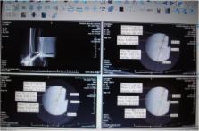
          Four different tissue phantoms namely hard tissue, soft tissue, air and water were scanned by three different CBCT and a CT system in two thicknesses (full and half) and two image acquisition settings (high and low kVp and mA). The images were analyzed at three sites (middle, periphery and intermediate) using eFilm software. The difference in density values was analyzed by ANOVA and correction coefficient test (P<0.05).

          Results:

          There was a significant difference between density values in CBCT and CT scans in most situations, and CBCT values were not similar to CT values in any of the phantoms in different thicknesses and acquisition parameters or the three different sites. The correction coefficients confirmed the results.

          Conclusions:

          CBCT is not reliable for tissue density assessment. The results were not affected by changes in thickness, acquisition parameters or locations.

          Related collections

          Most cited references21

          • Record: found
          • Abstract: found
          • Article: not found

          Clinical applications of cone-beam computed tomography in dental practice.

          Cone-beam computed tomography (CBCT) systems have been designed for imaging hard tissues of the maxillofacial region. CBCT is capable of providing sub-millimetre resolution in images of high diagnostic quality, with short scanning times (10-70 seconds) and radiation dosages reportedly up to 15 times lower than those of conventional CT scans. Increasing availability of this technology provides the dental clinician with an imaging modality capable of providing a 3-dimensional representation of the maxillofacial skeleton with minimal distortion. This article provides an overview of currently available maxillofacial CBCT systems and reviews the specific application of various CBCT display modes to clinical dental practice.
            Bookmark
            • Record: found
            • Abstract: found
            • Article: not found

            CBCT-based bone quality assessment: are Hounsfield units applicable?

            CBCT is a widely applied imaging modality in dentistry. It enables the visualization of high-contrast structures of the oral region (bone, teeth, air cavities) at a high resolution. CBCT is now commonly used for the assessment of bone quality, primarily for pre-operative implant planning. Traditionally, bone quality parameters and classifications were primarily based on bone density, which could be estimated through the use of Hounsfield units derived from multidetector CT (MDCT) data sets. However, there are crucial differences between MDCT and CBCT, which complicates the use of quantitative gray values (GVs) for the latter. From experimental as well as clinical research, it can be seen that great variability of GVs can exist on CBCT images owing to various reasons that are inherently associated with this technique (i.e. the limited field size, relatively high amount of scattered radiation and limitations of currently applied reconstruction algorithms). Although attempts have been made to correct for GV variability, it can be postulated that the quantitative use of GVs in CBCT should be generally avoided at this time. In addition, recent research and clinical findings have shifted the paradigm of bone quality from a density-based analysis to a structural evaluation of the bone. The ever-improving image quality of CBCT allows it to display trabecular bone patterns, indicating that it may be possible to apply structural analysis methods that are commonly used in micro-CT and histology.
              Bookmark
              • Record: found
              • Abstract: found
              • Article: not found

              Deriving Hounsfield units using grey levels in cone beam computed tomography.

              an in vitro study was performed to investigate the relationship between grey levels in dental cone beam CT (CBCT) and Hounsfield units (HU) in CBCT scanners. a phantom containing 8 different materials of known composition and density was imaged with 11 different dental CBCT scanners and 2 medical CT scanners. The phantom was scanned under three conditions: phantom alone and phantom in a small and large water container. The reconstructed data were exported as Digital Imaging and Communications in Medicine (DICOM) and analysed with On Demand 3D(R) by Cybermed, Seoul, Korea. The relationship between grey levels and linear attenuation coefficients was investigated. it was demonstrated that a linear relationship between the grey levels and the attenuation coefficients of each of the materials exists at some "effective" energy. From the linear regression equation of the reference materials, attenuation coefficients were obtained for each of the materials and CT numbers in HU were derived using the standard equation. HU can be derived from the grey levels in dental CBCT scanners using linear attenuation coefficients as an intermediate step.
                Bookmark

                Author and article information

                Journal
                J Dent (Tehran)
                J Dent (Tehran)
                JOD
                JOD
                Journal of Dentistry (Tehran, Iran)
                Tehran University of Medical Sciences
                1735-2150
                2008-2185
                March 2016
                : 13
                : 2
                : 108-115
                Affiliations
                [1 ] Oral and Maxillofacial Radiologist, Tehran, Iran
                [2 ] Assistant Professor of Oral and Maxillofacial Radiology, Oral and Maxillofacial Radiology Department, Shahed University of Medical Sciences, Dental School, Tehran, Iran
                [3 ] Assistant Professor of Oral and Maxillofacial Radiology, Oral and Maxillofacial Radiology Department, Shahid Beheshti University of Medical Sciences, Dental School, Tehran, Iran
                Author notes
                [* ]Corresponding author: Z. Azizi, Department of Oral and Maxillofacial Radiology, Oral and Maxillofacial Radiology Department, Shahed University of Medical Sciences, Dental School, Tehran, Iran, dr_azizi_mp@ 123456yahoo.com
                Article
                jod-13-108
                5139928
                27928239
                b6f84883-5d2b-4422-b222-0ea7c88440a7
                Copyright© Dental Research Center, Tehran University of Medical Sciences

                This work is licensed under a Creative Commons Attribution-NonCommercial 3.0 Unported License which allows users to read, copy, distribute and make derivative works for non-commercial purposes from the material, as long as the author of the original work is cited properly.

                History
                : 27 July 2015
                : 08 January 2016
                Categories
                Original Article

                Dentistry
                bone density,cone-beam computed tomography,tomography, x-ray computed
                Dentistry
                bone density, cone-beam computed tomography, tomography, x-ray computed

                Comments

                Comment on this article