0
views
0
recommends
+1 Recommend
0 collections
    0
    shares
      • Record: found
      • Abstract: found
      • Article: found
      Is Open Access

      Loss of control eating in children is associated with altered cortical and subcortical brain structure

      research-article

      Read this article at

      Bookmark
          There is no author summary for this article yet. Authors can add summaries to their articles on ScienceOpen to make them more accessible to a non-specialist audience.

          Abstract

          Introduction

          Loss of control (LOC) eating is the perceived inability to control how much is eaten, regardless of actual amount consumed. Childhood LOC-eating is a risk factor for the development of binge-eating disorder (BED), but its neurobiological basis is poorly understood. Studies in children with BED have shown both increased gray matter volume in regions related to top-down cognitive control (e.g., dorsolateral prefrontal cortex) and reward-related decision making (e.g., orbital frontal cortex) relative to healthy controls. However, no studies have examined brain structure in children with LOC-eating. To identify potential neurobiological precursors of BED, we conducted secondary analysis of five studies that conducted T1 MPRAGE scans.

          Methods

          A total of 143, 7–12-year-old children ( M = 8.9 years, 70 boys) were included in the study, 26% of which ( n = 37) reported LOC-eating (semi-structured interview). Age, sex, and obesity status did not differ by LOC-eating. Differences between children with and without LOC were examined for gray matter volume, cortical thickness, gyrification, sulci depth, and cortical complexity after adjusting for age, sex, total intercranial volume, weight status, and study.

          Results

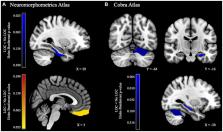
          Children with LOC, relative to those without, had greater gray matter volume in right orbital frontal cortex but lower gray matter volume in right parahippocampal gyrus, left CA4/dentate gyrus, and left cerebellar lobule VI. While there were no differences in cortical thickness or gyrification, children with LOC-eating had great sulci depth in left anterior cingulate cortex and cuneus and greater cortical complexity in right insular cortex.

          Discussion

          Together, this indicates that children with LOC-eating have structural differences in regions related to cognitive control, reward-related decision-making, and regulation of eating behaviors.

          Related collections

          Most cited references74

          • Record: found
          • Abstract: found
          • Article: not found

          An automated labeling system for subdividing the human cerebral cortex on MRI scans into gyral based regions of interest.

          In this study, we have assessed the validity and reliability of an automated labeling system that we have developed for subdividing the human cerebral cortex on magnetic resonance images into gyral based regions of interest (ROIs). Using a dataset of 40 MRI scans we manually identified 34 cortical ROIs in each of the individual hemispheres. This information was then encoded in the form of an atlas that was utilized to automatically label ROIs. To examine the validity, as well as the intra- and inter-rater reliability of the automated system, we used both intraclass correlation coefficients (ICC), and a new method known as mean distance maps, to assess the degree of mismatch between the manual and the automated sets of ROIs. When compared with the manual ROIs, the automated ROIs were highly accurate, with an average ICC of 0.835 across all of the ROIs, and a mean distance error of less than 1 mm. Intra- and inter-rater comparisons yielded little to no difference between the sets of ROIs. These findings suggest that the automated method we have developed for subdividing the human cerebral cortex into standard gyral-based neuroanatomical regions is both anatomically valid and reliable. This method may be useful for both morphometric and functional studies of the cerebral cortex as well as for clinical investigations aimed at tracking the evolution of disease-induced changes over time, including clinical trials in which MRI-based measures are used to examine response to treatment.
            Bookmark
            • Record: found
            • Abstract: not found
            • Article: not found

            MatchIt: Nonparametric Preprocessing for Parametric Causal Inference

              Bookmark
              • Record: found
              • Abstract: found
              • Article: not found

              A dual-networks architecture of top-down control.

              Complex systems ensure resilience through multiple controllers acting at rapid and slower timescales. The need for efficient information flow through complex systems encourages small-world network structures. On the basis of these principles, a group of regions associated with top-down control was examined. Functional magnetic resonance imaging showed that each region had a specific combination of control signals; resting-state functional connectivity grouped the regions into distinct 'fronto-parietal' and 'cingulo-opercular' components. The fronto-parietal component seems to initiate and adjust control; the cingulo-opercular component provides stable 'set-maintenance' over entire task epochs. Graph analysis showed dense local connections within components and weaker 'long-range' connections between components, suggesting a small-world architecture. The control systems of the brain seem to embody the principles of complex systems, encouraging resilient performance.
                Bookmark

                Author and article information

                Contributors
                Journal
                Front Psychol
                Front Psychol
                Front. Psychol.
                Frontiers in Psychology
                Frontiers Media S.A.
                1664-1078
                11 January 2024
                2023
                : 14
                : 1237591
                Affiliations
                [1] 1Department of Nutritional Science, The Pennsylvania State University , University Park, PA, United States
                [2] 2Division of Endocrinology, Diabetes, and Metabolism, Children's Hospital Los Angeles , Los Angeles, CA, United States
                [3] 3Department of Health and Life Sciences, Florida State University , Tallahassee, FL, United States
                [4] 4United States Department of Agriculture , Washington, DC, United States
                [5] 5Department of Food Science, The Pennsylvania State University , University Park, PA, United States
                Author notes

                Edited by: Filip Morys, McGill University Health Centre, Canada

                Reviewed by: Grace Shearrer, University of Wyoming, United States; Roberta Dalle Molle, Douglas Mental Health University Institute, Canada

                *Correspondence: Alaina L. Pearce, azp271@ 123456psu.edu
                Article
                10.3389/fpsyg.2023.1237591
                10808807
                38274697
                b6c6bf31-fdab-4f9c-82a6-a4a5d29bec80
                Copyright © 2024 Pearce, Fuchs, Adise, Masterson, Fearnbach, English and Keller.

                This is an open-access article distributed under the terms of the Creative Commons Attribution License (CC BY). The use, distribution or reproduction in other forums is permitted, provided the original author(s) and the copyright owner(s) are credited and that the original publication in this journal is cited, in accordance with accepted academic practice. No use, distribution or reproduction is permitted which does not comply with these terms.

                History
                : 27 June 2023
                : 20 December 2023
                Page count
                Figures: 2, Tables: 2, Equations: 0, References: 76, Pages: 9, Words: 7664
                Funding
                This work was supported by the National Institutes of Health (NIH), NIDDK [F32 DK122669-01, R01 DK110060, K01 DK135847], the USDA [PEN 04565 and 2011–67001-30117], The Pennsylvania State University’s Social, Life, & Engineering Sciences Imaging Center’s [UL1TR000127], and NCATS [TR002015, UL1 TR002014, and UL1TR000127].
                Categories
                Psychology
                Original Research
                Custom metadata
                Pediatric Psychology

                Clinical Psychology & Psychiatry
                loss of control-eating,gray matter volume,surface morphology,children,mri

                Comments

                Comment on this article